Rhizophagus intraradices
(reference accession UT126)
Whole Spores | ||
|---|---|---|
|
|
|
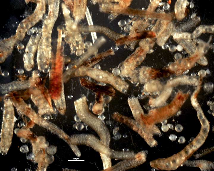
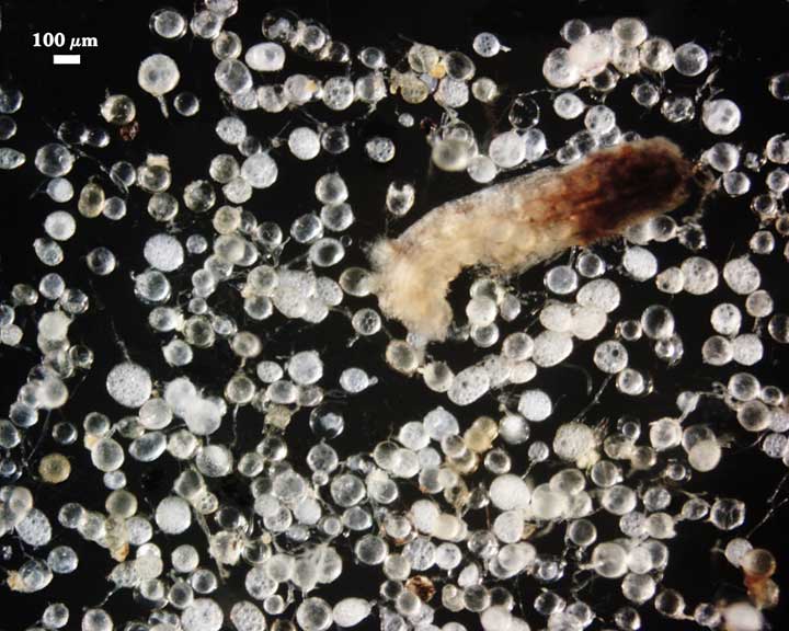
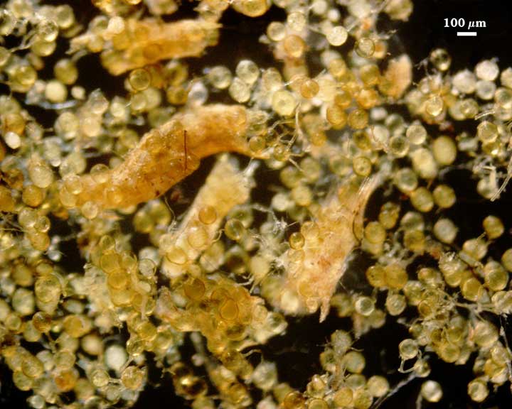
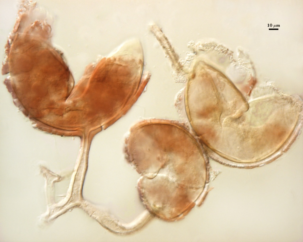
 COLOR: White, pale cream (0-0-10-0) to yellow brown (0-10-40-0), sometimes with a green tint. Color is highly variable within this continuum.
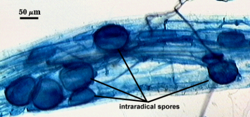
SHAPE: Globose, subglobose, irregular, with many spores elliptical (especially those extracted from within mycorrhizal roots).
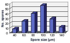
SIZE DISTRIBUTION: 40-140 µm, mean = 93.3 µm (n = 170).
Subcellular Structure of Spores
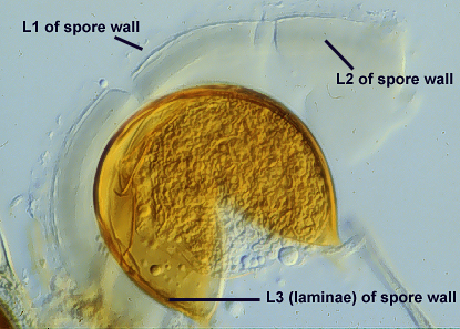
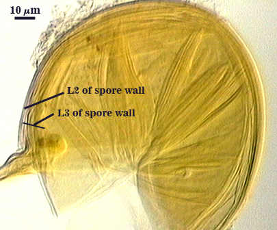
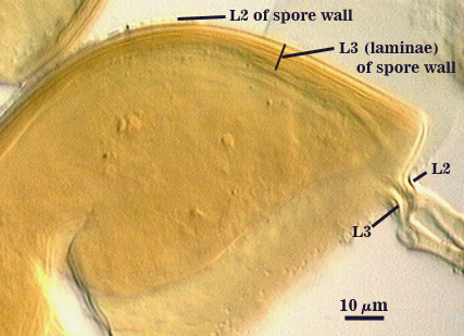
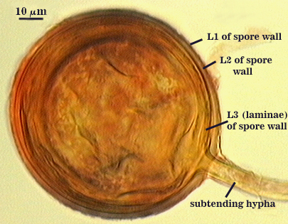
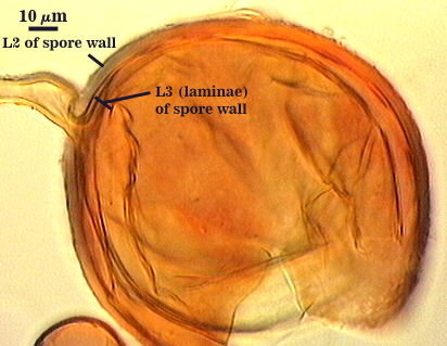
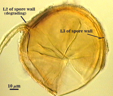
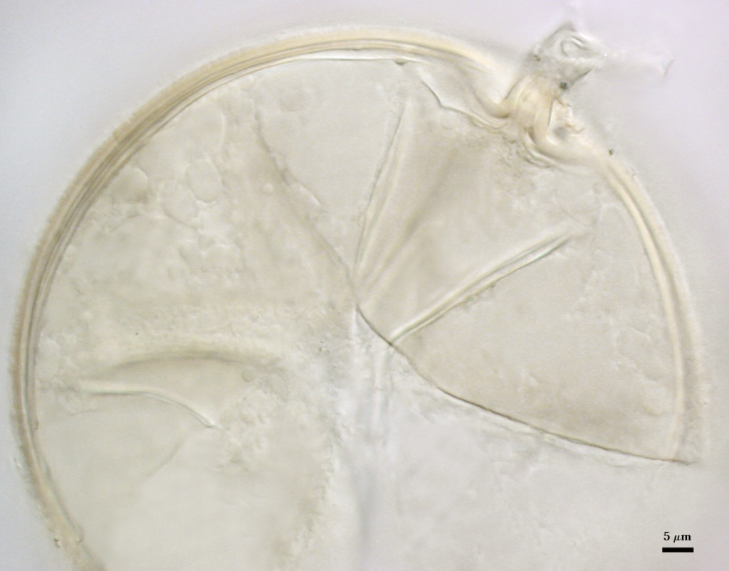
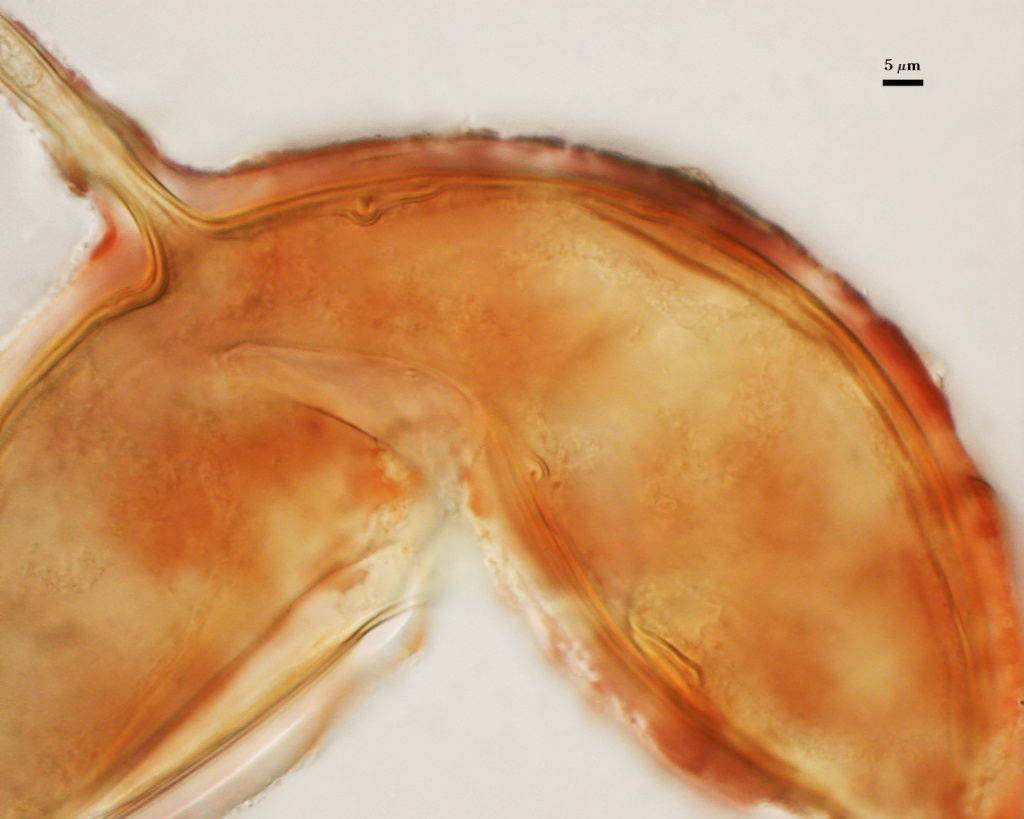
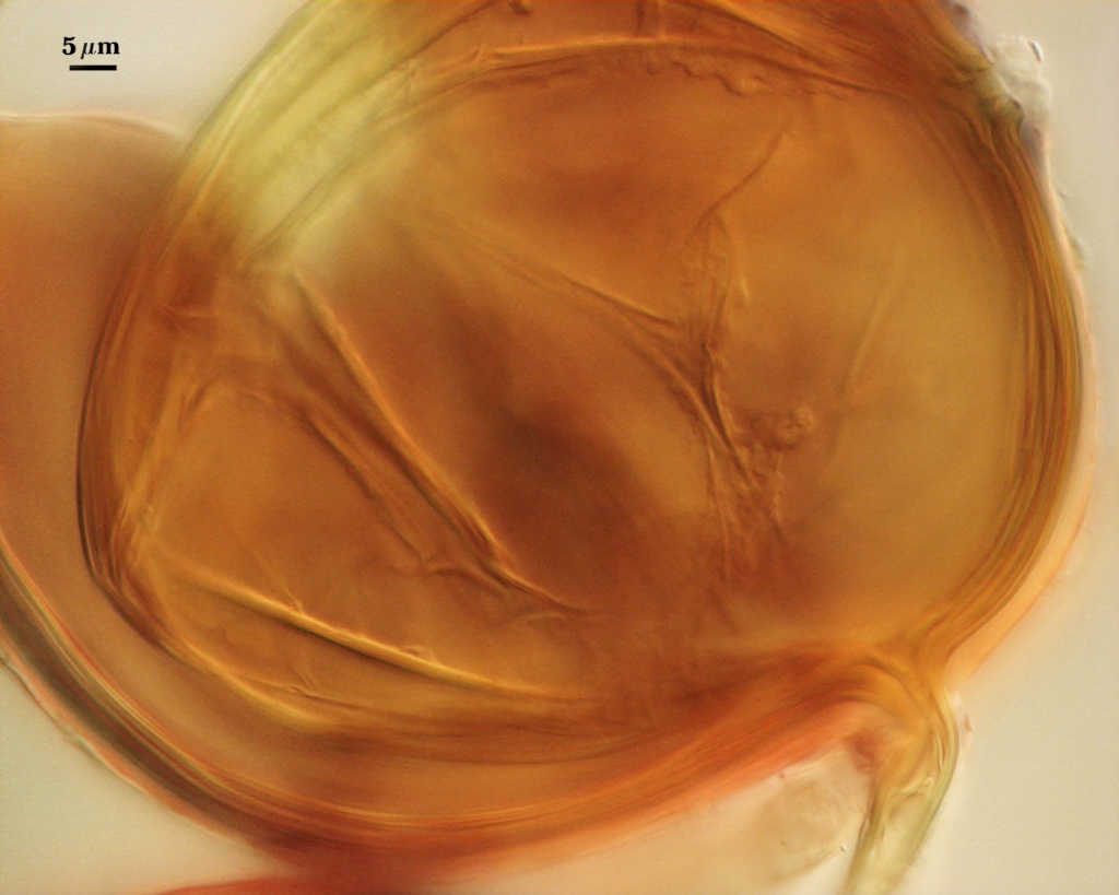
SPORE WALL: Three layers (L1, L2 and L3), with only the first layer present in juvenile spores and subtending hyphal wall (so they are colorless); L2 and L3 then formed sequentially in both the spore wall and in the wall of the subtending hypha (see sequence of photos below, from left to right).
| L2 and L3 | |||
|---|---|---|---|
|
|
|
|
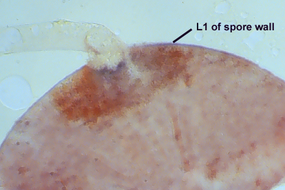
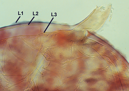
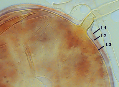
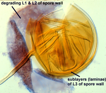
L1: Outermost layer, hyaline, mucilagenous, 0.6-3.2 µm thick, staining pinkish red (0-60-30-10) to pale purple (20-60-20-10) in Melzer’s reagent when intact in juvenile spores. With age, this layer almost always degrades and decomposes naturally and from the action of microorganisms, after which it appears granular and may accumulate some debris.
L2: Adherent to the mucilagenous outer layer, hyaline, 1.5-4.9 µm thick (mean of 2.6 µm) when intact in young spores. With age, this layer degrades concomitant with L1 and also acquires a granular appearance or sloughs in patches. Mature spores often lack both L1 and L2 or they are present together as rough patches.
L3: A layer in that it consists of pale yellow-brown (0-0-80-0 to 5-0-80-0) sublayers (or laminae) that either remain adherent or separate with applied pressure. Degree of separation among sublayers varies considerably among spores and often is affected by age, amount of parasitism, or amount of applied pressure after mounting. In juvenile spores, initial sublayer(s) are 0.5-1 µm and the layer thicken with formation of additional sublayers. Thickness varies from 3.2-12 µm (mean = 7.2 µm) in mature spores. This layer forms simultaneously in the wall of the subtending hypha.
| In PVLG | ||
|---|---|---|
|
|
|
| In PVLG and Melzer's reagent (1:1 v/v) | ||
|---|---|---|
|
|
|
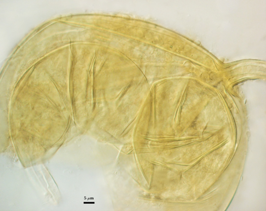
Subtending Hypha
SHAPE: Cylindrical to slightly flared, occasionally slightly constricted (see photos above).
WIDTH: 11-18 µm (mean = 15 µm).
COMPOSITE WALL THICKNESS: 3.2-6.4 µm.
WALL STRUCTURE: Three layers (L1, L2 and L3) that are continuous with the three layers of the spore wall. The two outer layers are the only ones present in early stages of spore formation, and both thin or degrade with spore maturation. Hyphae attached to spores often react lightly in Melzer’s reagent, indicating some residual material of L1. Sublayers of L3 also can separate along the hyphal length, although they are fewer in number.
OCCLUSION: Innermost sublayer(s) of L3 of the spore wall which do not extend into the subtending hypha. Occasionally a plug partitions spore from hyphal contents.
Germination
A germ tube emerges from the lumen of the subtending hypha. It appears to arise from the innermost sublayer of L3. Some specimens show germ tubes arising from broken ends of hyphal fragments some distance from the spore by the same mechanism. This behavior could account for the high infectivity of hyphal fragments of fungi in this species.
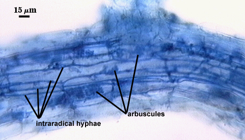
Mycorrhizae
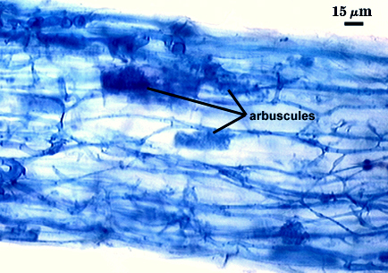
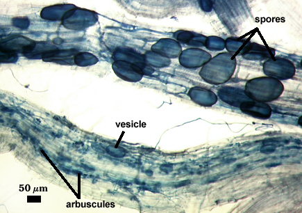
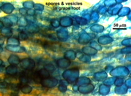
Arbuscules and hyphae in cortical cells of sorghum, sudangrass, corn and red clover roots stain darkly in trypan blue. Numerous vesicles (or spores) often form near entry points along with the arbuscule-hyphal network. It is uncertain as the extent intraradical vesicles are still able to further differentiate into spores, because they can co-occur in roots.
| Arbuscules in cortical cells of corn | ||
|---|---|---|
|
|
|
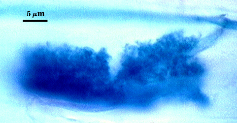
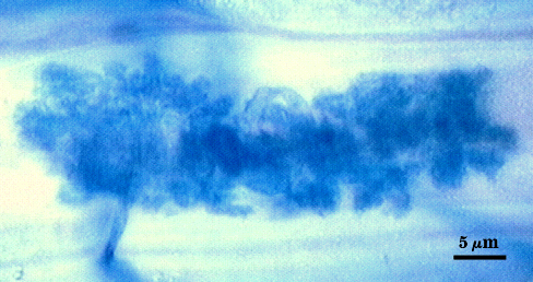
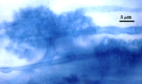
| Mycorrhizal structures in corn | ||
|---|---|---|
|
|
|
| Spores in corn pot culture | Spores field-grown apple |
|---|---|
|
|
Notes
Many species reported in the literature as Glomus fasciculatum appear now to be synonymous with G. intraradices. One report (Biermann and Linderman, 1983) did not recognize the vesicle/spore relationship of G. intraradices and reported vesicles to be infective.
Spore size and color are highly variable not only within one population of the species, but also between isolates. Amount of yellow color is a function of thickness of L3 in the spore wall, and smaller spores will naturally have a thinner layer. Some isolates may consist mostly of smaller spores (and thus be mostly hyaline, subhyaline to pale yellow) or larger spores (and thus be mostly a dark yellow to yellow brown).
Many cultures which were started from spores diagnosed as Glomus aggregatum subsequently produced typical spores of R. intraradices, so a continuum exists in number of sublayers in the laminate layer of the spore wall (possibly related to size) that suggests these two species are synonymous.
The images below can be uploaded into your browser by clicking on the thumbnail or can be downloaded to your computer by clicking on the link below each image. Please do not use these images for other than personal use without expressed permission from INVAM.
High Resolution Images | |
|---|---|
 |  |
 |  |
|  |
 |  |
Reference
- Biermann, B. and R. G. Linderman. 1983. Use of vesicular-arbuscular mycorrhizal roots, intraradical vesicles, and extraradical vesicles as inoculum. New Phytologist 95: 97-105.