Claroideoglomus lamellosum
(reference accession ON393)
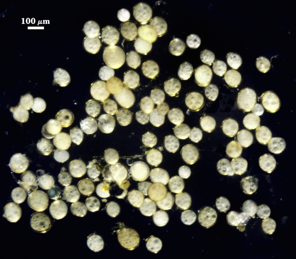
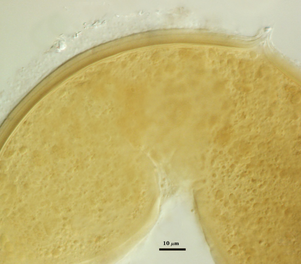
Whole Spores
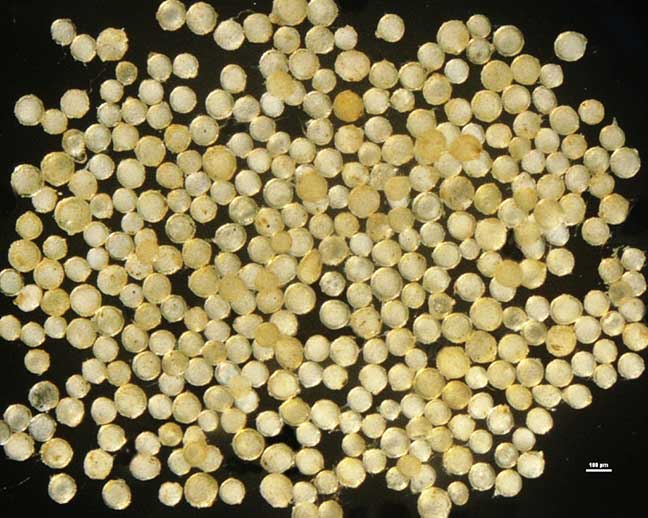
 COLOR: Cream (0-0-10-0) to pale yellow (0-0-40-0)
SHAPE: Globose to subglobose
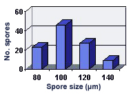
SIZE DISTRIBUTION: 80-140 µm, mean = 104 µm (n = 105)
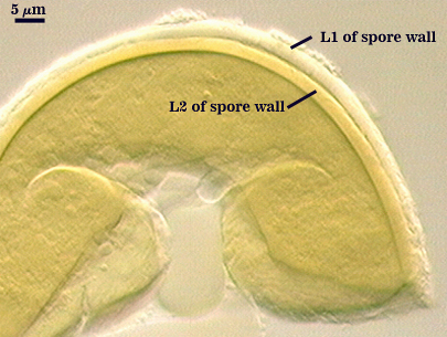
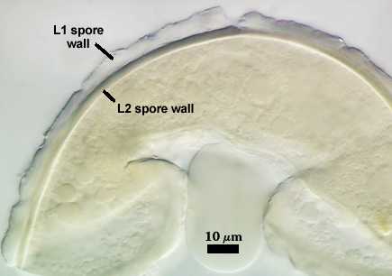
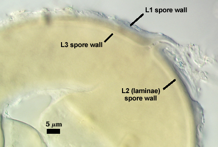
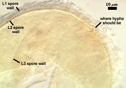
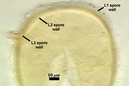
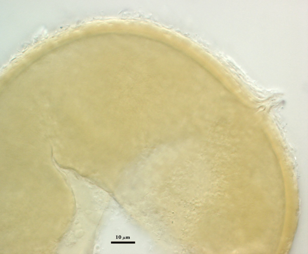
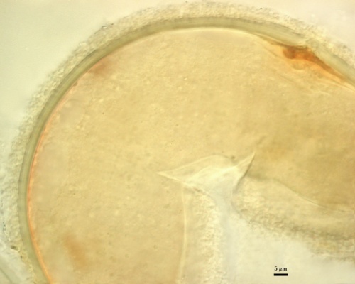
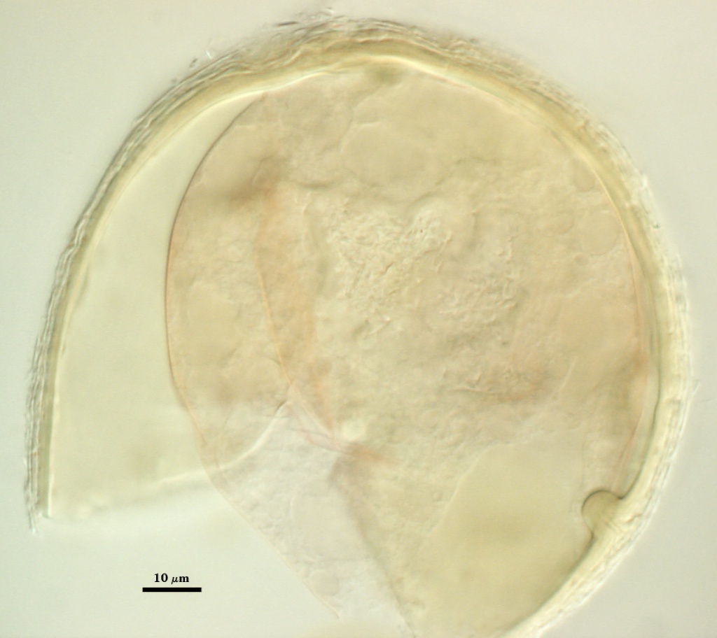
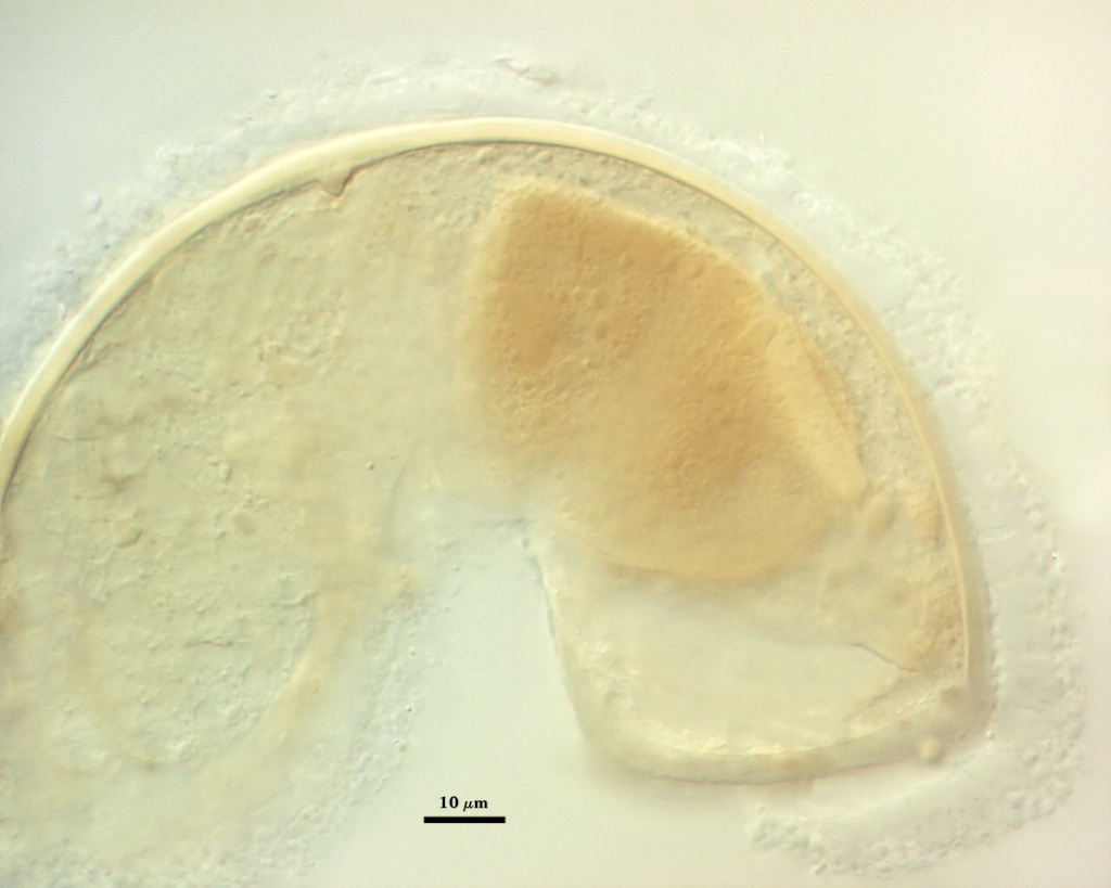
Subcellular Structure of Spores
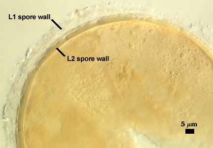
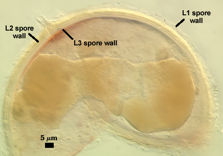
SPORE WALL: Three layers (L1, L2, and L3), with all persisting in mature spores.
L1: Hyaline to light yellow (0-0-20-0) layer, 4-14 µm thick; producing no reaction in Melzer’s reagent. This layer tends to degrade on the surface, giving it a flaky appearance.
L2: A layer consisting of thin and tightly adherent pale yellow (0-0-20-0) to slightly darker yellow (0-0-40-0) sublayers (or laminae), 6-12 µm thick in mature spores, losing pigmentation abruptly in the subtending hyphal wall (a continuation of the spore wall layer).
L3: A thin layer concolorous with the laminate layer, < 1 µm thick, producing folds when it is very thin. Its appearance closely resembles that of a flexible inner wall (=”membranous” wall), but it extends into the subtending hypha as part of the hyphal wall. It is considered a separate layer rather than a sublayer of L3 because it consistently separates to varying degrees from the spore wall in at least 80% of spores in populations of all accessions. When adherent to L3, it cannot be seen. In some spores, this layer stains faint pink in Melzer’s reagent (left two photos in bottom row below), a rare occurrence among Glomus species.
| In PVLG | |||
|---|---|---|---|
|
|
|
|

| In PVLG and Melzer's Reagent | |||
|---|---|---|---|
|
|
|
|
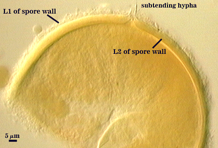
Subtending Hypha
SHAPE: Cylindrical to slightly flared, occasionally recurved slightly.
WIDTH: 8-13 µm wide at spore base.
COMPOSITE WALL THICKNESS: < 1-2 µm.
WALL STRUCTURE: Two layers (L1 and L2) continuous with the two layers of the spore wall.
L1: Two layers (L1 and L2) continuous with the outer two layers of the spore wall, 1.2-2.4 µm thick in the region of attachment, tapering within 15 µm of the spore to less than 1.0 µm thick (only L2 present). In some spores, the hyphal wall is so fragile that it breaks from the spore and cannot be seen.
OCCLUSION: The innermost layer of the spore wall (L3), a recurved “septum” which grows from the wall of the subtending hyphae, or the two structures together.
Germination
A germ tube emerges from the lumen of the subtending hypha.
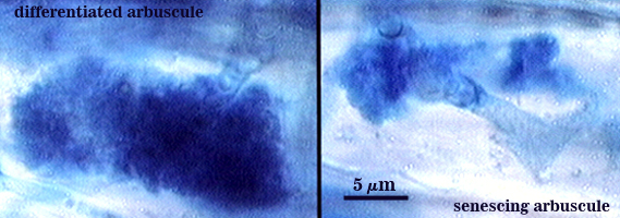
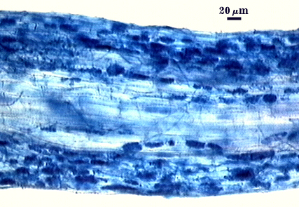
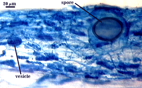
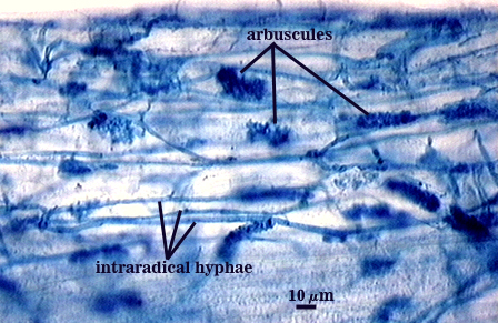
Mycorrhizae
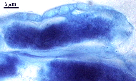
Source host for this analysis was corn (Zea mays L.). Arbuscules branch from intraradical hyphae 2.5-4 µm in diameter, staining darkly in trypan blue when fully formed and functional. Branch hyphae from the main trunk of the arbuscule narrow incrementally to numerous fine tips.
Intraradical hyphae of a mycorrhiza also stain darkly in trypan blue. They tend to grow intercellularly between cortical cells, with the hyphal network consisting of numerous “H and h” branches and occasional coils. Vesicle development is sporadic and patchy, rarely abundant in any particular infection unit.
| Arbuscules in corn roots | ||
|---|---|---|
|
| |
| All mycorrhizal structures in corn roots | ||
|---|---|---|
|
|
|
Notes
This fungus is not known outside sandy habitats of the Great Lakes region. Dalpe et al. (1992) found the species in dune grass in Georgian Bay, Ontario, Canada and Bailey’s Harbor, Wisconsin. This particular accession was from Wasaga Beach, Ontario (provided by Yolande Dalpe).
Walker and Vestberg (1998) had some concern about the extent of similarities between this species and Glomus claroideum, but decided to maintain it as a distinct taxon. We agree with that assessment.
The images below can be uploaded into your browser by clicking on the thumbnail or can be downloaded to your computer by clicking on the link below each image. Please do not use these images for other than personal use without expressed permission from INVAM.
High Resolution Images | |
|---|---|
 |  |
 |  |
 |  |
Links to Gene Sequences in Genbank
Reference
- Dalpe, Y., R. E. Koske, and L. L. Tews. 1992. Glomus lamellosum sp. nov.: A new Glomaceae associated with beach grass. Mycotaxon 63:289-293.
- Walker, C. and M. Vestberg. 1998. Synonymy amongst the arbuscular mycorrhizal fungi: Glomus claroideum, G. maculosum, G. multisubstensum and G. fistulosum. Annals of Botany 82:601-624.