Acaulospora rehmii
(reference accession BR237)
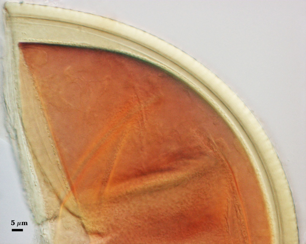
Whole Spores
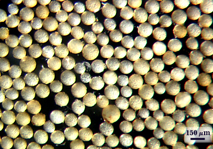
 COLOR: Yellow-brown (0-10-40-0) to orange-brown (0-30-100-0), most dark yellow-brown (0-20-60-0).
SHAPE: Globose, subglobose, occasionally irregular.
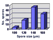
SIZE DISTRIBUTION: 100-160 µm, mean = 141 µm (n = 101).
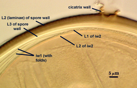
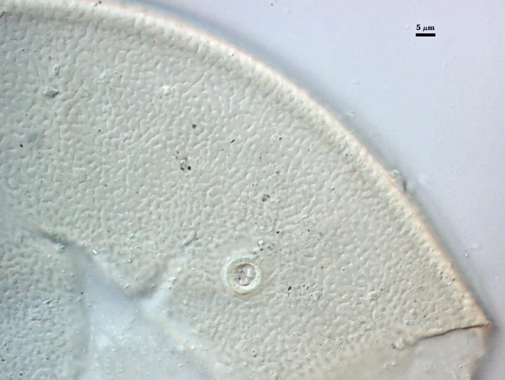
Subcellular Structure of Spores
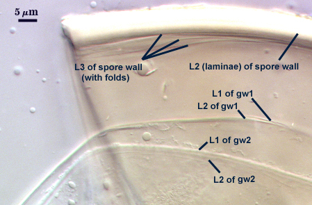
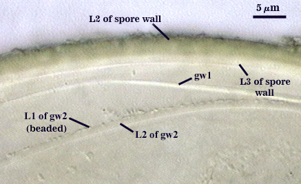
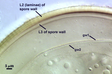
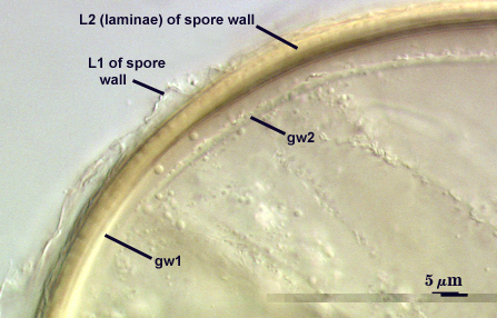
SPORE WALL: Three layers (L1, L2, and L3), with L1 continuous with the wall of the neck of the parent sporiferous saccule and the latter two layers being synthesized with development of the spore.
L1: Hyaline; 0.8-1.2 µm thick; degrading and sloughing early in spore wall differentiation so that it is usually absent from the surface of mature spores (or only sporadic patches remain).
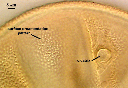
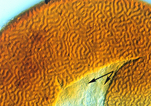
L2: A layer that thickens initially by formation of pale yellow (0-0-20-0) to yellow-brown (0-20-60-0) sublayers (or laminae), with the outer sublayers forming depressions < 0.5-0.8 µm wide and 0.5-1.4 µm deep that create a network of ridges organized in a complex labyrinthian pattern when viewed from the spore surface. At maturity, the pore between spore and saccule neck is bridged by sublayers of this layer to form an “endospore”.
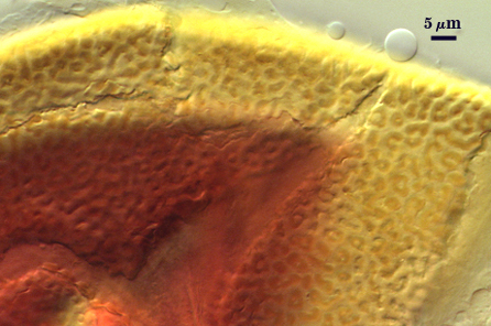
L3: L2 appears (photo above) to consist of two “zones”, with an inner zone of sublayers that separates collectively or only the innermost sublayer separates and appears as a distinct structure that resembles a flexible inner wall (which has caused considerable confusion in defining subcellular organization of these spores). For the moment, we are interpreting this zone as a separate layer.
| Spores mounted in PVLG | |||
|---|---|---|---|
|
|
|
|
| Spores mounted in PVLG and Melzer’s reagent (1:1 v/v) | |||
|---|---|---|---|
|
|
|
|
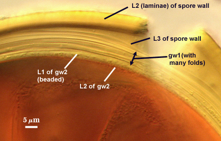
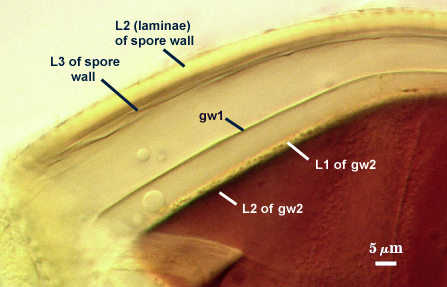
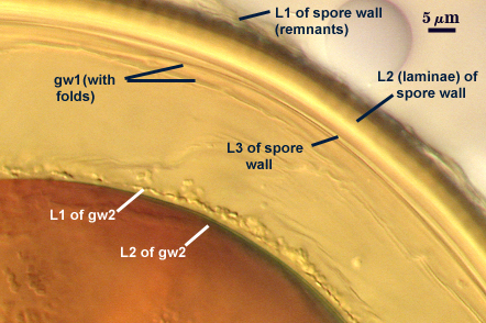
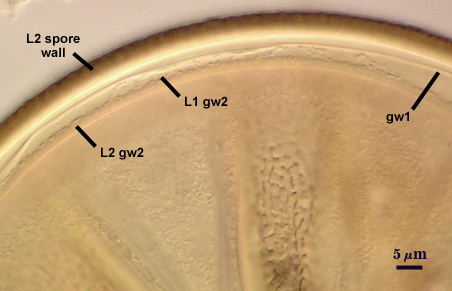
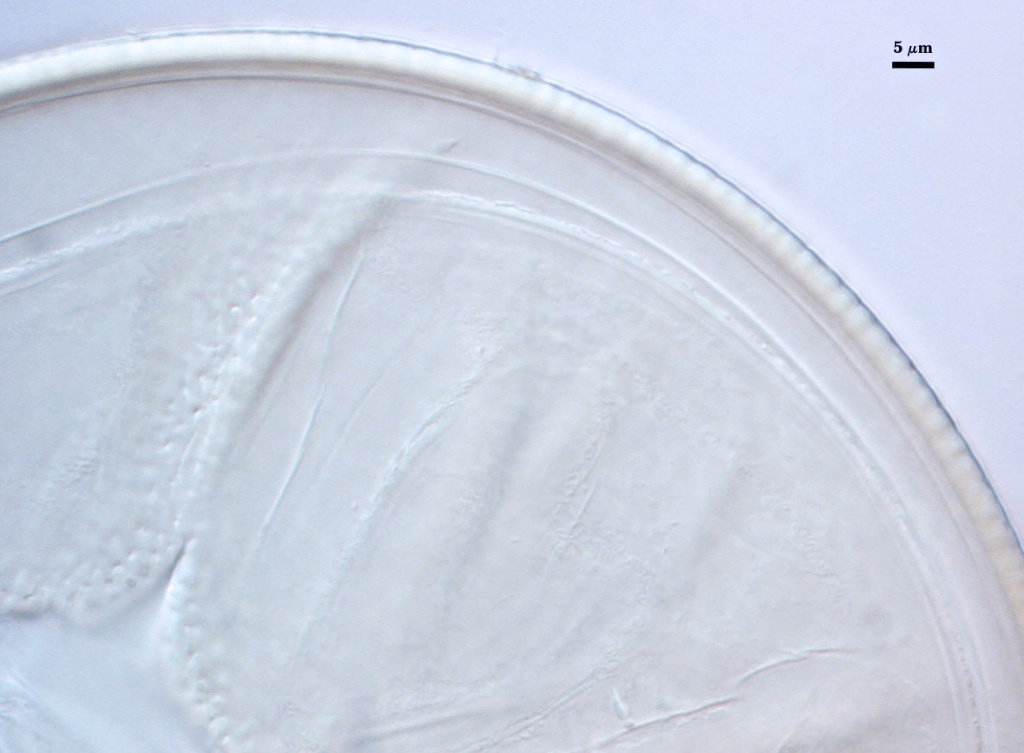
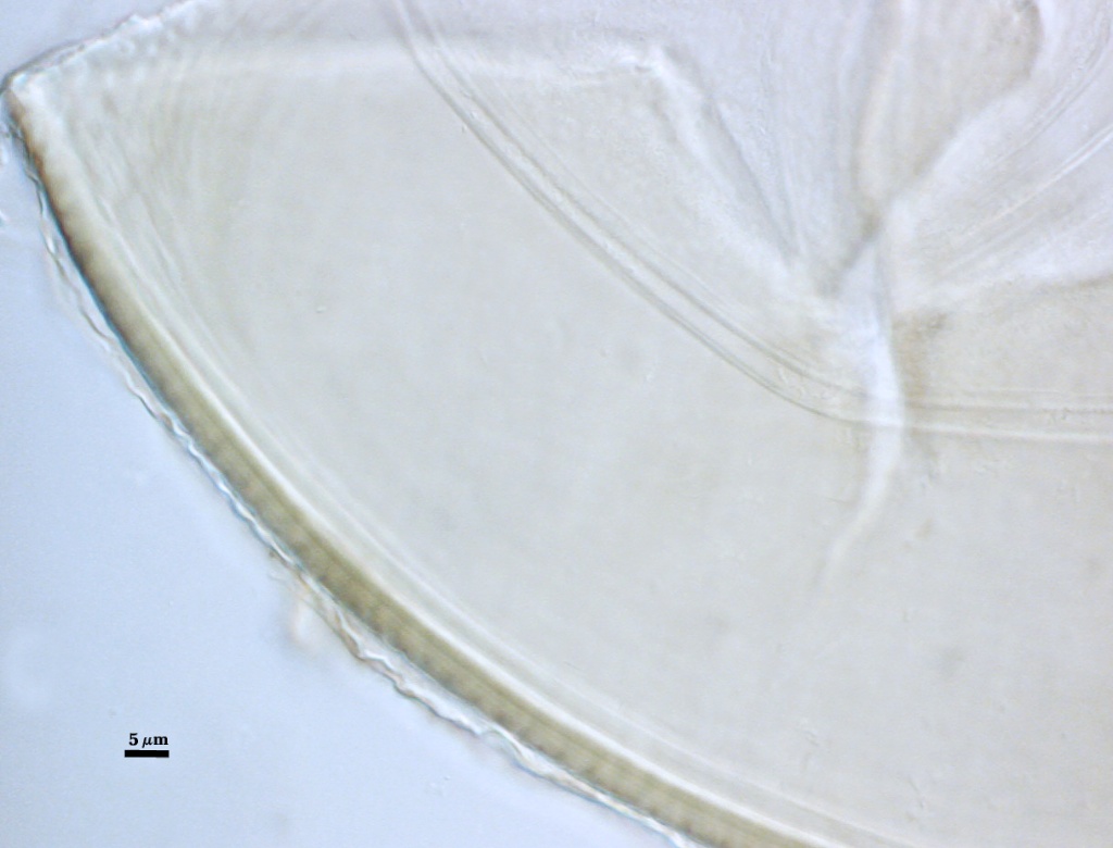
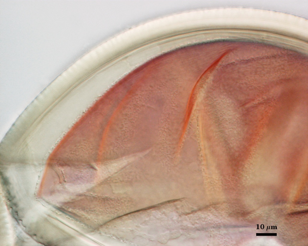
FLEXIBLE GERMINAL WALLS: Two inner walls (gw1 and gw2) can be seen in all spores IF they separate when a spore is broken. 9 GW1: Consisting of two thin hyaline layers, each < 0.5 µm thick, which often are tightly adherent (and thus appearing as a single layer). This wall usually separates readily from the spore wall, although it can be hard to distinguish from folds of L3 (or inner sublayers of L2) when they separate (see far right photo above). Neither layer produces any visible reaction in Melzer’s reagent.
GW1: N/A
GW2: Consisting of two, usually adherent, hyaline layers formed (L1 and L2). L1 is 0.7-1.2 µm thick, with granular excresences (or “beads”) that tend to become dislodged and float away with applied pressure. These “beads” are stabilized after preservation in formalin, but otherwise may be absent on mounted spores within a few months of storage. L2 is 0.6-1.0 µm thick (measured in PVLG where boundaries can be seen) and appears pale reddish-orange (0-40-60-0) to a reddish-brown (20-80-80-0) in Melzer’s reagent. This color reaction is due to observing L2 through layers of the spore wall. When separated completely from the spore wall, L2 stains light pink (0-30-20-0) to pinkish-red (0-60-50-10).
Cicatrix
A circular to ovoid scar indicating region of contact between spore and saccule neck during spore synthesis; the depression within the scar is ridges, 7.0-12.9 µm in diameter (mean = 9.5 µm).
| Spore Growth | |
|---|---|
 |  |
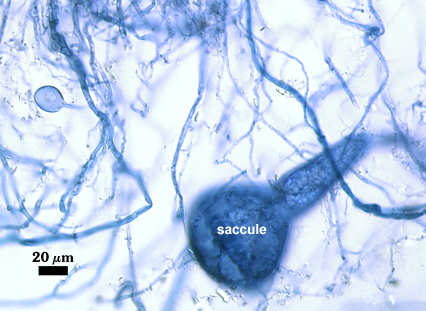
Sporiferous Saccule
COLOR: Hyaline
SHAPE: Globose to subglobose.
SIZE: 90-140 µm, mean = 129 µm.
SACCULE WALL: One hyaline layer, smooth surface, 1.2-2.0 µm thick.
DISTANCE 40-90 µm.
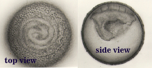
Germination
An ovoid “germination orb” forms on the innermost germinal wall (gw2), from which germ tubes form and penetrate through the spore wall. This orb is difficult to see except in older spores where contents have cleared with fusion of lipid globules in the spore lumen, mostly because it is wide enough to span most of the diameter of the spore and so edges of the orb are seen only with a limited range of spore orientations. Appears to resemble that described by Spain (1992) for A. scrobiculata. The photos above were provided courtesy of Joyce Spain.
Notes
Germinal wall structure is difficult to see and interpret until spores are completely cleared (slides stored for at least 30 days) because of the light-scattering caused by ornamentations in the laminate layer (L2) of the spore wall. Holotype specimens of this species (photo at right) often were so parasitized that inner walls were missing and obvious colonization by other fungi could be seen in the lumen of spores. Therefore, it is not surprising that the first inner wall and the beaded surface of gw2 were not mentioned in the species description (Spain, 1992) and spores were described as being much darker (red-brown to black). Dimensions of ridges and depressions of the labyrinthian surface ornamentations of L2 of the spore wall were described by Sieverding and Toro (1987) as being much larger than those observed in this isolate (ridges 1.0-5.0 µm high and depressions 1.0-4.5 µm wide).
The images below can be uploaded into your browser by clicking on the thumbnail or can be downloaded to your computer by clicking on the link below each image. Please do not use these images for other than personal use without expressed permission from INVAM.
High Resolution Images | ||
|---|---|---|
 |  |  |
 |  | |
Reference
- Spain, J. L. 1992. Patency of shields in water-mounted spores of four species in Acaulosporaceae (Glomales). Mycotaxon 43:331-339.
- Sieverding, E. and S. Toro T. 1987. Acaulospora denticulata sp. nov. and Acaulospora rehmii sp. nov. (Endogonaceae) with ornamented spore walls. Angewandte Botanik 61:217-223.