Dentiscutata rubra
(reference accession BR211)
Whole Spores
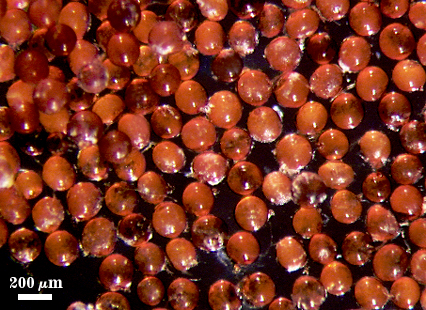
 COLOR: Dark orange-brown (0-60-100-0) to red-brown (20-80-100-0), most tending toward the latter at maturity. Immature spores are white to cream with a rose tint (0-10-40-0) under a dissecting microscope.
SHAPE: Globose to subglobose.
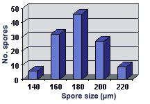
SIZE DISTRIBUTION: 140-220 µm, mean = 180 µm (n = 114).
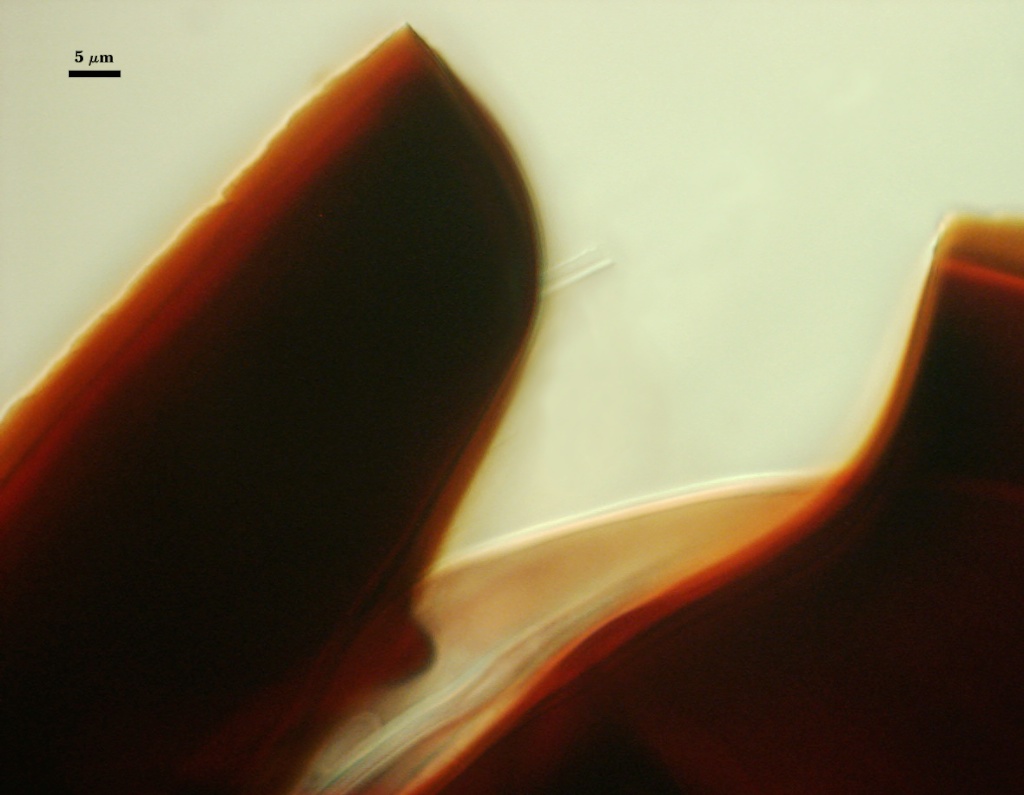
Subcellular Structure of Spores
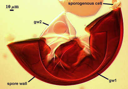
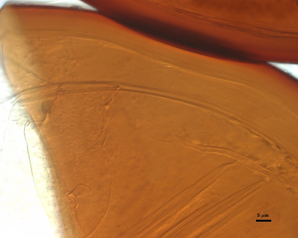
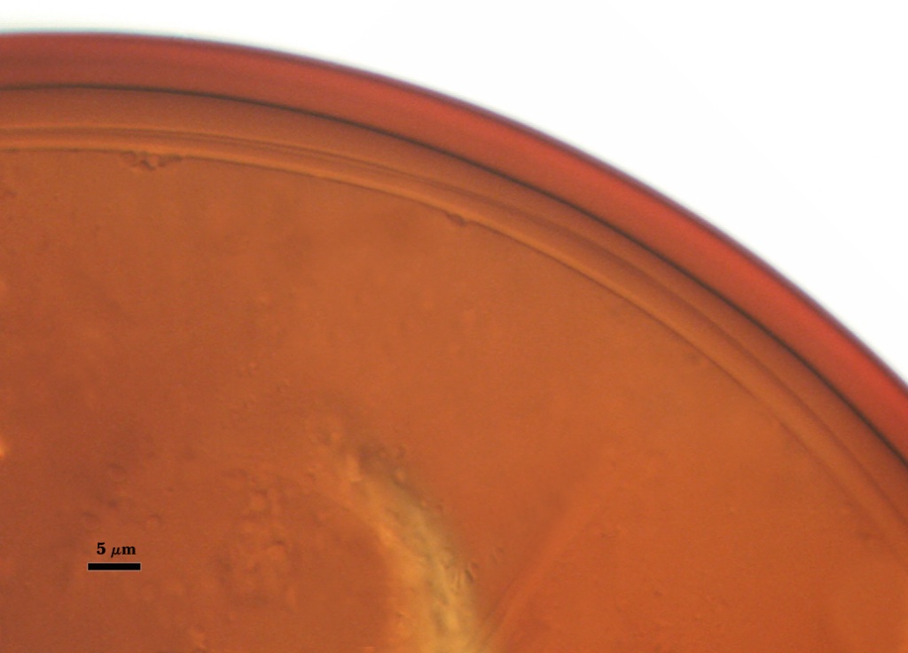
SPORE WALL: Three layers (L1, L2, and L3) with L1 and L2 of equal thickness at the start of spore differentiation, with L2 then becoming somewhat plastic as it thickens. It is uncertain whether L3 forms concurrent with or following differentiation of L2 because it is so hard to resolve even in mature spores.
| Sequence in the differentiation of subcellular structure (left to right) | |||
|---|---|---|---|
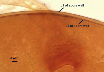 | 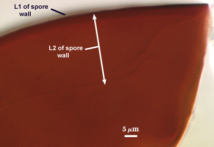 |  |  |
L1: An outer smooth permanent rigid layer, pale brown (0-20-50-10) in color; produces no reaction in Melzer’s reagent.
L2: A layer consisting of fine orange-brown (20-80-80-0) to red-brown (20-80-100-0) sublayers (or laminae); 3.5-12 µm thick in mature spores (and depending on amount of pressure applied to a coverslip). In water (a medium in which this layer is rigid, thickness ranges from 3.5-7.5 µm. This layer stains dark red-black (20-80-70-10) in Melzer’s reagent.
L3: A very thin hyaline flexible layer, < 1 µm thick, can be seen more clearly in vigorously crushed spores, but usually only where it attaches to the spore wall near the occluding plug and the sporogenous cell region (pictured in structure of subtending hypha below). A homologous layer was first detected in S. cerradensis, subsequently in S. heterogama, and provided the impetus to search for a similar layer in other Scutellospora species.
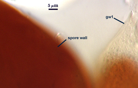
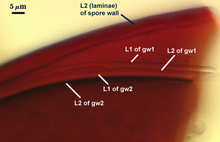
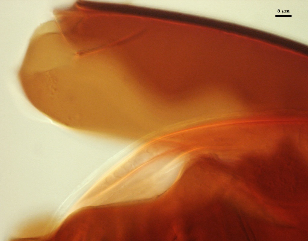
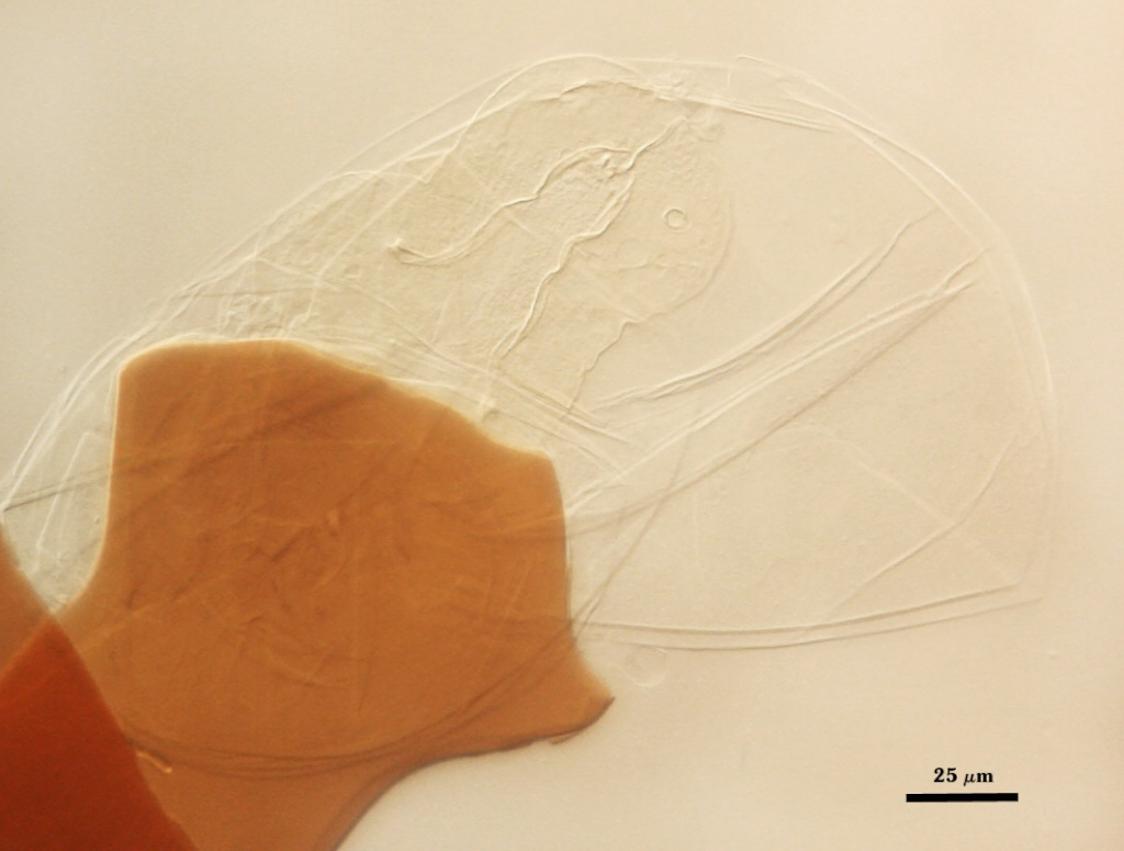
GERMINAL WALLS: Two bilayered hyaline flexible inner walls (gw1 and gw2) are synthesized consecutively after the spore wall has completes differentiation. They are formed completely separate from the spore wall. In many mature spores, both germinal walls tend to stay closely paired, even with applied pressure (see photos below), so that together, they appears a a single germinal wall with 2-4 layers. Differential interference contrast optic are needed to clearly resolve layers of both germinal walls.
| In PVLG | In PVLG and Melzer's reagent | |
|---|---|---|
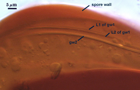 |  | 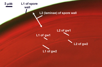 |
GW1: Two layers are formed (L1 and L2) that are tightly adherent. L1 is less than 0.5 µm thick; L2 is slightly thicker (0.6-0.8 µm). Composite wall thickness is 0.6-1.1 µm. Both layers are thin enough that when tightly adherent, they appear as one layer.
GW2: Two layers are formed (L1 and L2) that are tightly adherent. L1 is 0.5-0.6 µm thick; L2 is 0.6-1.4 µm thick. Both of these layers also are thin enough to appear as one layer in PVLG. However, only L2 differentially stains a pale pinkish-purple (0-20-20-0) to slightly darker pinkish-purple (20-40-20-0) in Melzer’s reagent.
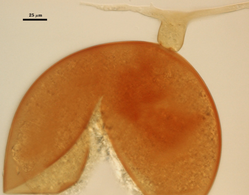
Subtending Hypha
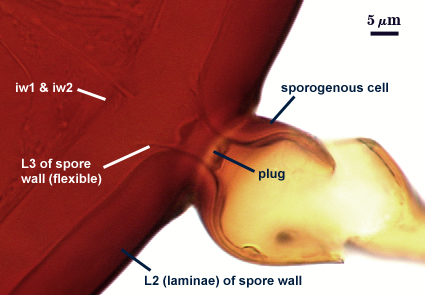
WIDTH OF SPOROGENOUS CELL: 18-35 µm.
SPOROGENOUS CELL WALL: Two layers (L1 and L2) probably are present (continuous with the two layers of the spore wall), but only L2 is readily discernible at the level of the compound microscope.
L2: Dark orange-brown in color (20-80-80-0), 1.2-3 µm thick near the spore and then thinning to 0.6-0.8 µm beyond the sporogenous cell.
OCCLUSION: Closure by a plug concolorous with L2 of the spore wall.
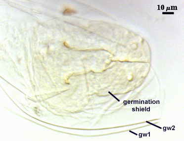
Germination
COLOR: Pale yellow-brown (0-30-70-10) to darker orange-brown (0-40-100-10).
SHAPE: Oblong, violin-shaped, pale yellow-brown (0-5-20-0), dimensions of a60-68×82-109 µm. Margin of the shields is fairly smooth, with only a few lobes and attendant paired germ holes. Position of the shield is on iw2.
Auxiliary Cells
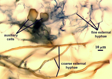
Aggregate (2-11) cells borne on coiled dark brown (0-40-80-0) hyphae 2.5-5 µm in diameter. Each cell is; thin-walled (< 1 µm thick), with a light brown (0-20-80-10) wall in transmitted light; cells almost smooth with some having slightly raised but flattened knobs on the surface.
Mycorrhizae
Extraradical hyphae of two types: one thinner, 0.8-1.6 µm wide, and hyaline; the other thicker, 2.4-5 µm wide, and yellow-brown (0-40-100-0). The latter often is more abundant near entry points. Appressoria from these hyphae also are yellow-brown (see below). Intraradical arbuscules and hyphae consistently stain darkly in roots treated with trypan blue. Arbuscules with many fine terminal branches from a swollen trunk, 1.6-7.9 µm wide. Hyphae of variable thickness, ranging from 1.6-6.3 µm wide, usually as a consequence of irregular-shaped swellings. They grow parallel o the root axis or form loos to tight coils in the cortex. Hyphal entry ponts often occurred through root hairs, but also through root epidermal cell walls.
| Arbuscules in corn roots | |
|---|---|
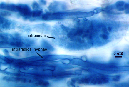 | 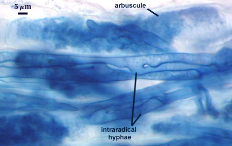 |
| Mycorrhizae with entry points | |
|---|---|
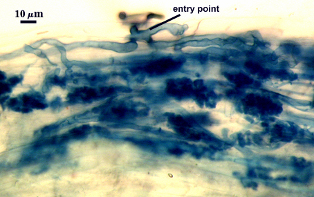 | 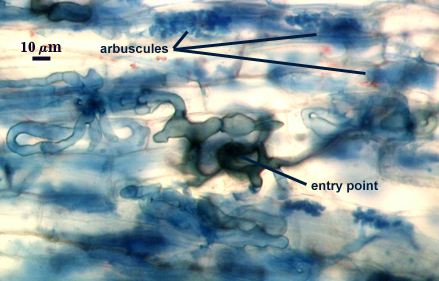 |
Notes
This species is indistinguishable morphologically from D. heterogama, except that the outer layer (L1) of the spore wall has no ornmanentations in D. rubra. This pattern of morphological variation (in layers of the spore wall) is consistent with species level differences in all other genera. The transition stage during differentiation of the laminate layer (L2) is much more obvious in D. heterogama than D. rubra. In the former, the L2 layer is quite plastic when hyaline, and becoming more rigid as pigmentation is acquired. In the latter, the hyaline state is much less frequent (suggesting that it is of shorter duration and hence likely to be sampled at any given time) and plasticity is not as pronounced.
The images below can be uploaded into your browser by clicking on the thumbnail or can be downloaded to your computer by clicking on the link below each image. Please do not use these images for other than personal use without expressed permission from INVAM.
High Resolution Images | |
|---|---|
 |  |
 |  |
 |  |
Reference
- Stürmer, S. L. and J. B. Morton. 1999. Scutellospora rubra, a new arbuscular mycorrhizal species from Brazil. _Mycological Research. 103:949-954.