Dentiscutata heterogama
(reference accession IL203)
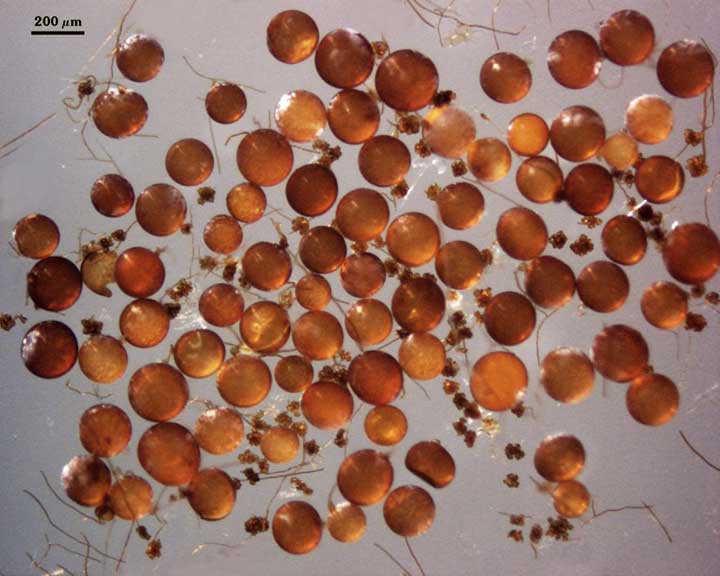
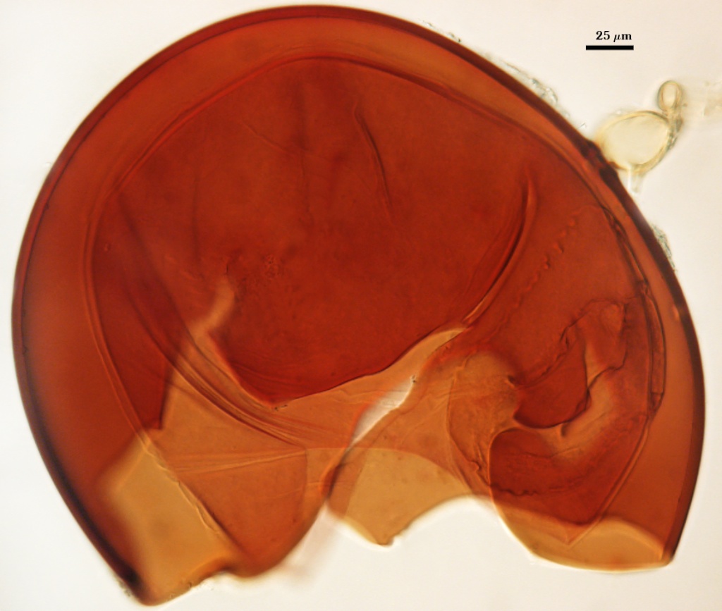
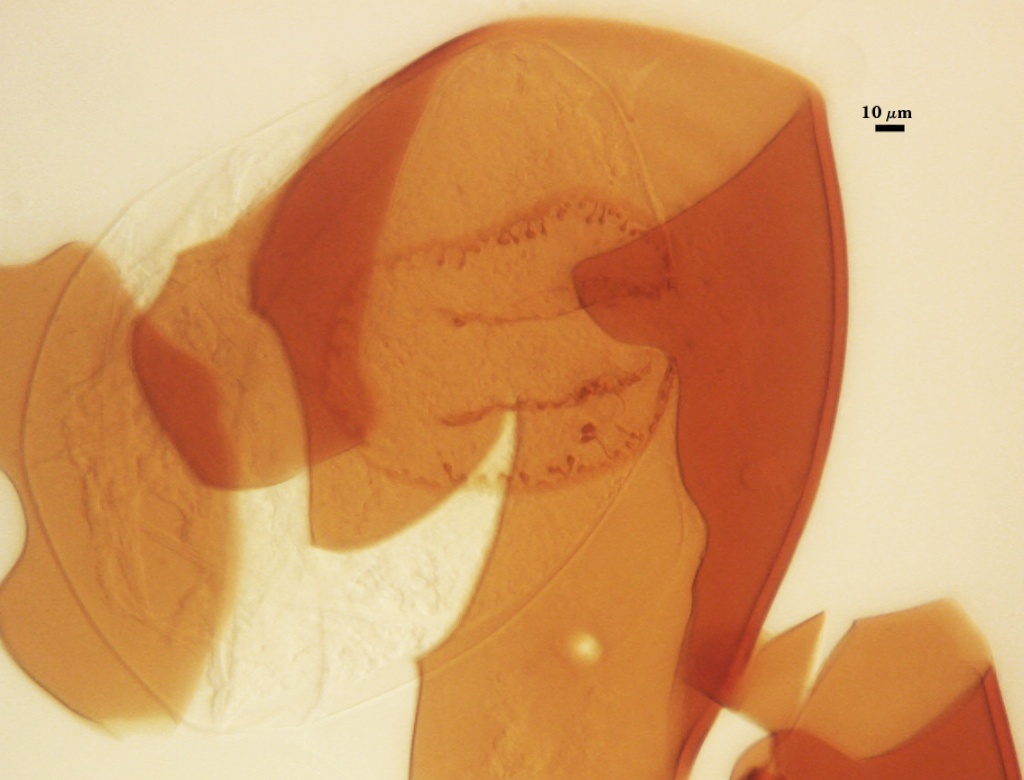
Whole Spores
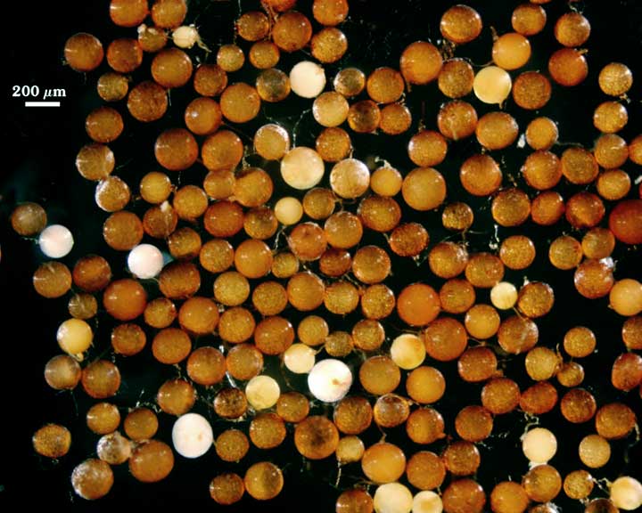
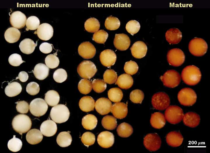
 COLOR: Dark orange-brown (0-60-100-10) to red-brown (40-80-100-0). Immature spores are white to cream with a rose tint (0-10-40-0) and dense contents under a dissecting microscope and in water. If the young spores are placed in Melzer’s reagent, they will turn black because the immature spore wall is amorphous and stains a dark red-purple (see below).
SHAPE: Globose, subglobose to oblong or ellipsoid.
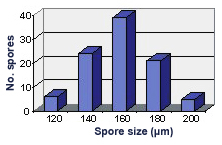
SIZE DISTRIBUTION: 120-200 µm, mean = 159 µm (n = 95).
Subcellular Structure of Spores
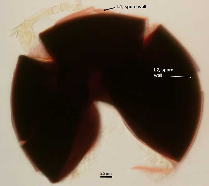
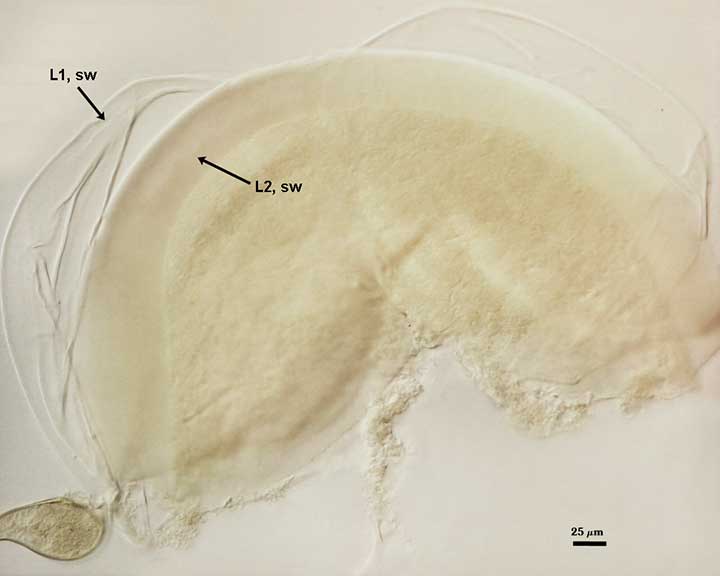
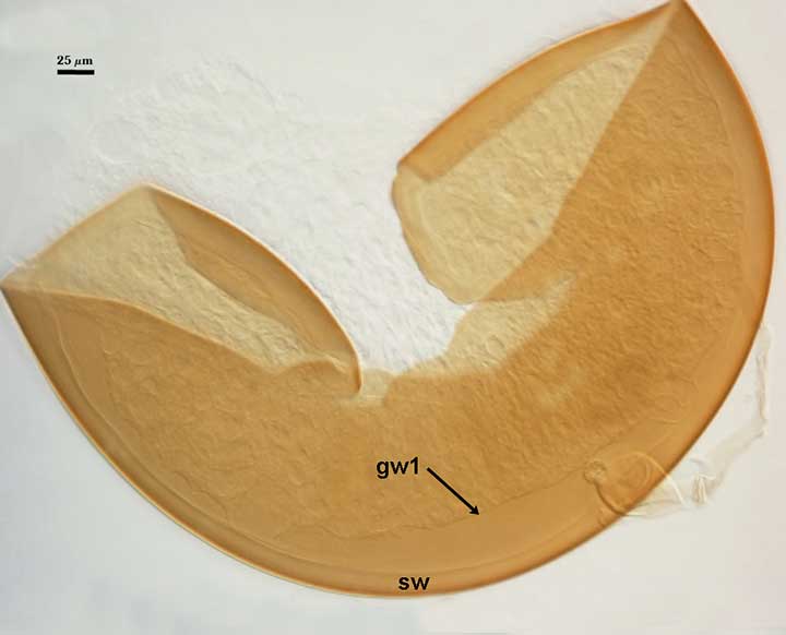
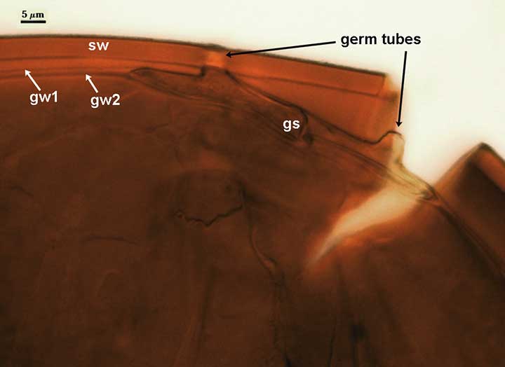
SPORE WALL: Three layers (L1, L2, and L3) with the middle layer (L2) undergoing a dramatic transformation from the juvenile to the mature state. This transformation is expressed in the transition in spore color. The sequence in the differentiation of this layer is chronicled in the photograph sequence below (from left to right). It is uncertain whether L3 forms concurrent with or following differentiation of L2 because it is so hard to resolve even in mature spores. This ontogenetic sequence was resolved by Morton and Msiska (2010) to reference against ontogeny of a stable albino mutant.
| Transition in color | L2 becomes amorphous, in Melzer’s | L2 amorphous, no stain | |
|---|---|---|---|
 |  |  |  |
| L2 condenses, becomes pigmented, gw1 forms | GW2 forms, then germ shield – in Melzer’s | ||
|---|---|---|---|
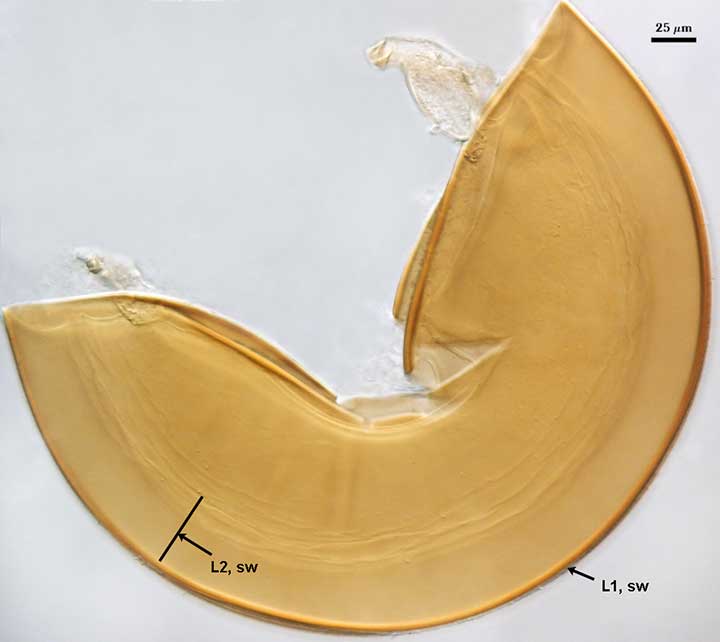 |  | 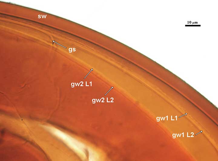 |  |
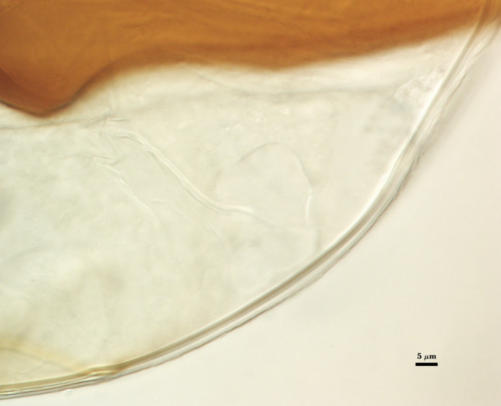
L1: An outer permanent rigid layer with tightly packed short rounded warts 1.0-2.5 µm high, pale brown (0-20-50-10) in color. Warts often become invisible in PVLG mountants after several months storage, although this effect is variable. They still are evident on the spore wall of holotype specimens.
L2: A layer consisting of fine orange-brown (20-80-80-0) to red-brown (20-80-100-0) sublayers (or laminae); 5.0-9.0 µm thick (mean = 7.4 µm) in mature spores. This layer stains dark red-brown (20-80-70-10) in Melzer’s reagent.
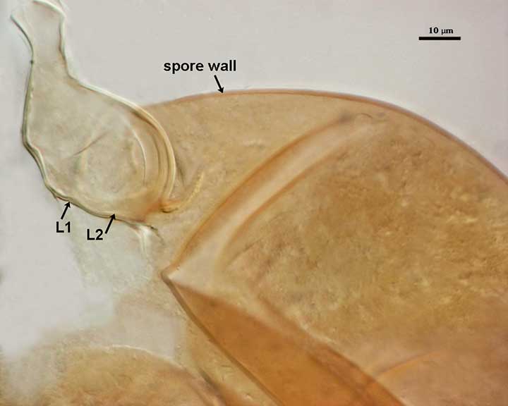
L3: A very thin hyaline flexible layer, < 1 µm thick, can be seen in vigorously crushed spores, usually only where it attaches to the spore wall near the occluding plug and the sporogenous cell region (not shown). A homologous layer was first detected in S. cerradensis and provided the impetus to search for a similar layer in other Scutellospora species.
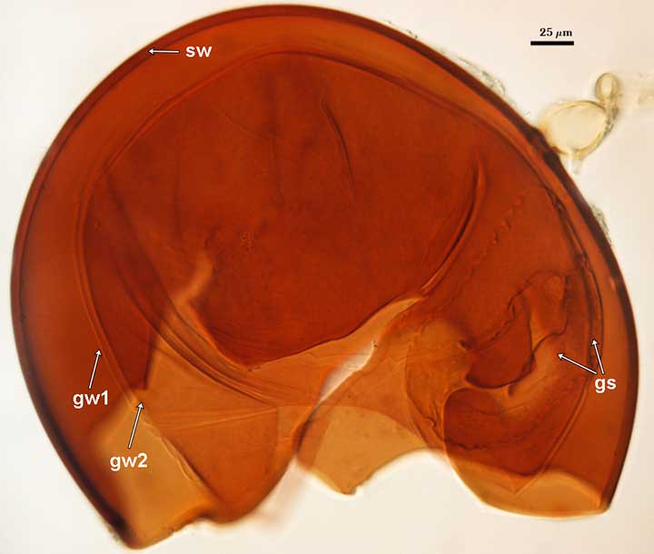
GERMINAL WALLS: Two bilayered flexible hyaline inner walls (gw1 and gw2) that are synthesized consecutively after the spore wall has completed differentiation. They are formed completely separate from the spore wall. In many mature spores, both germinal walls tend to stay closely paired, even with applied pressure (see photos below).
GW1: Two layers are formed (L1 and L2) that are tightly adherent. L1 is less than 0.5 µm thick; L2 is slightly thicker (0.8-1.5 µm). Both layers are thin enough that when tightly adherent, they appear as one layer. However, sometimes L1 separates in patches and resembles “warts” or undulations in the wall, much like that found in the only germinal wall of R. coralloidea, R. fulgida, R. gregaria, R. persica, and R. verrucosa.
GW2: Two layers are formed (L1 and L2) that are tightly adherent. L1 is 0.5-0.8 µm thick; L2 is 0.9-1.8 µm thick. Both of these layers also are thin enough to appear as one layer in PVLG. However, only L2 differentially stains a pinkish purple (0-60-30-0) to slightly darker purple (20-60-20-0) in Melzer’s reagent.
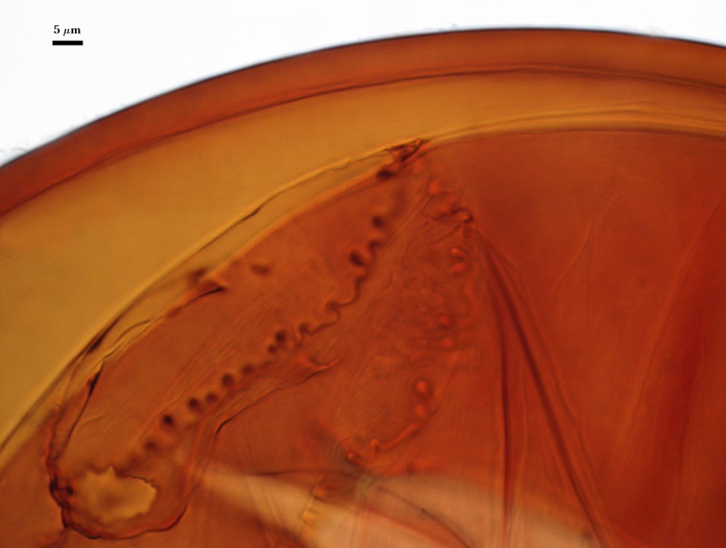
Subtending Hypha
WIDTH OF SPOROGENOUS CELL: 24-28 µm (mean = 26.4 µm) (see photos above).
SPOROGENOUS CELL WALL STRUCTURE: Two layers (L1 and L2) probably are present (continuous with the two layers of the spore wall), but only L2 is readily discernible at the level of the compound microscope.
L2: Orange-brown in color (0-60-80-0), 1.8-2.2 µm thick near the spore and then thinning to 0.6-0.8 µm beyond the sporogenous cell.
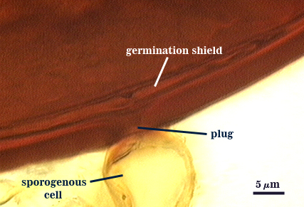
OCCLUSION: Closure by a plug concolorous with L2 of the spore wall.
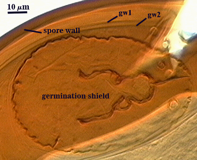
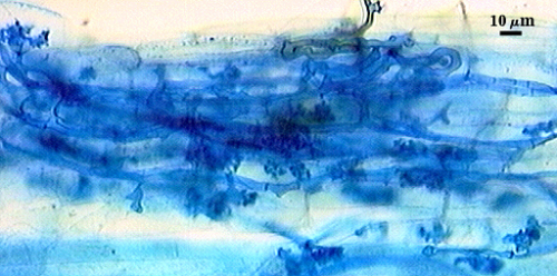
Germination
| Germination shield | |
|---|---|
 |  |
COLOR: Pale yellow-brown (0-30-70-10) to darker orange-brown (0-40-100-10).
SHAPE: Oblong, with length approximately 1.5 times that of the width. Margin of the shields is fairly smooth, with only a few folds and attendant paired germ holes. Shape of the shield resembles that of a violin. Position of the shield is on iw2.
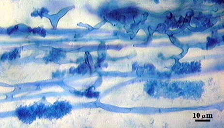
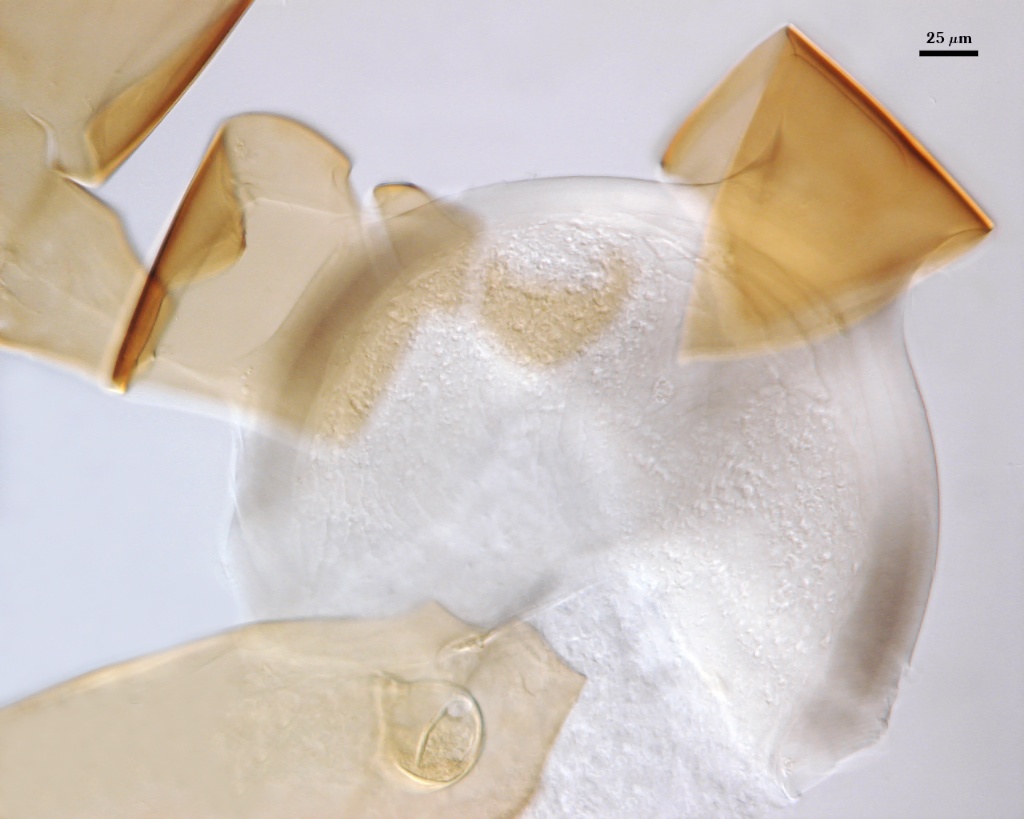
Auxiliary Cells
| Germination shield | |
|---|---|
 | 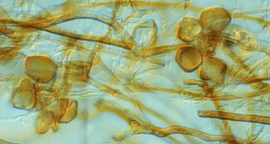 |
Aggregate (1-10) cells borne on coiled brown (20-40-80-0) hyphae 4-6 µm in diameter; thin-walled (< 1 µm thick), with a light brown (0-20-70-10) wall in transmitted light; cells almost smooth with some having slight undulations on the surface.
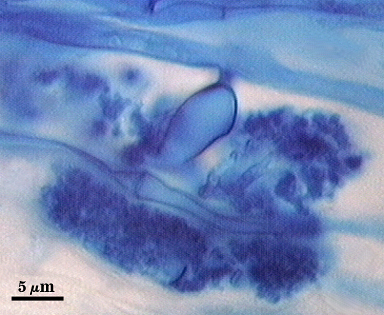
Mycorrhizae
Intraradical arbuscules and hyphae consistently stain darkly in roots treated with trypan blue. Arbuscules with many fine tips from a swollen trunk (see photos below). Hyphae often with knobs or projections, usually densely coiled near entry points.
| Arbuscules in corn | ||
|---|---|---|
 | 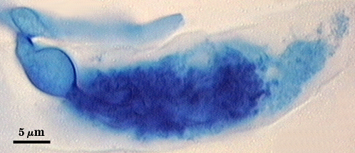 | 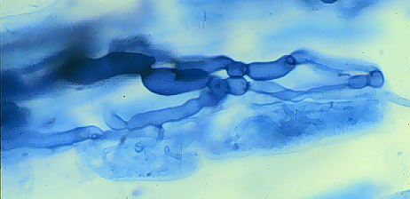 |
| Other mycorrhizal structures in corn | |
|---|---|
 |  |
Notes
The first germinal wall was redescribed (Koske and Walker, 1985) as having two “membranous walls” with some undefined adhesive causing them to stick together. The spores studied were neither fresh nor healthy, so the component structures were not recognized as two separate walls with two layers to each wall. Clarification occurred with a close examination of spore ontogeny and the relationship between inner walls and the position of the germination shield (Morton and Msiska, 2010).
The images below can be uploaded into your browser by clicking on the thumbnail or can be downloaded to your computer by clicking on the link below each image. Please do not use these images for other than personal use without expressed permission from INVAM.
High Resolution Images | |
|---|---|
|  |
 |  |
| |
 |  |
Links to Gene Sequences in Genbank
Reference
- Koske, R. E. and C. Walker. 1985. Species of Gigaspora (Endogonaceae) with roughened outer walls. Mycologia 77:702-720.
- Morton, J. B. and Z. Msiska. 2010. Ontogeny and phylogeny of a Scutellospora heterogama mutant, with implications for species variation in Glomeromycota. Fungal Biology 114:410-420.