Scutellospora calospora
(reference accession AU221)
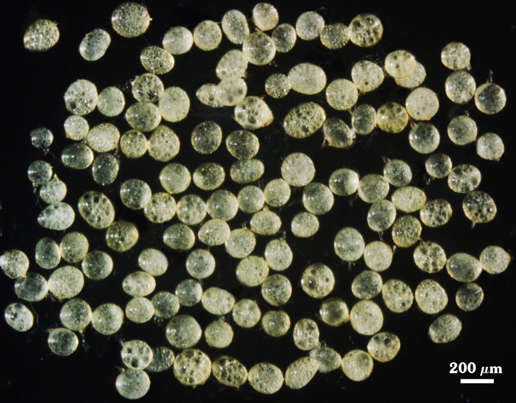
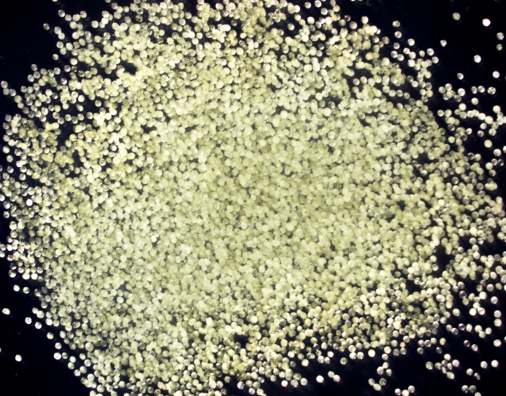
Whole Spores | ||
|---|---|---|
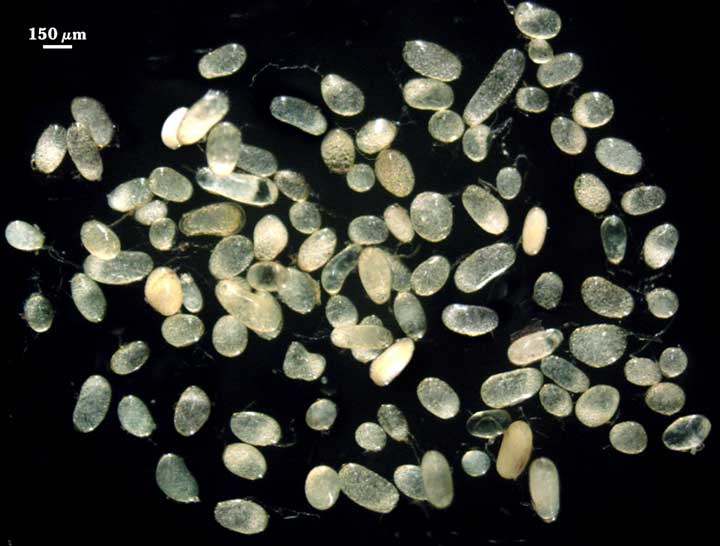 |  |  |
 COLOR: Pale yellow with a greenish tint (5-0-20-0) to yellow-brown with greenish tint (5-0-100-10) in older spores.
SHAPE: Wide range, from subglobose to ellipsoid to oblong, sometimes irregular. We have one strain from West Virginia that is exclusively subglobose and another one from Poland that has been exclusively ellipsoid for more than five propagation cycles.
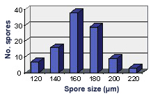
SIZE DISTRIBUTION: 120-220 µm, mean = 165 µm (n = 100).
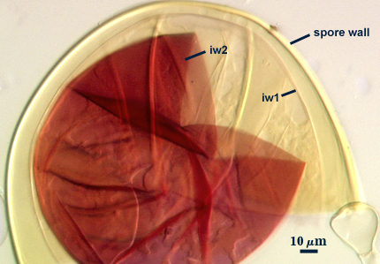
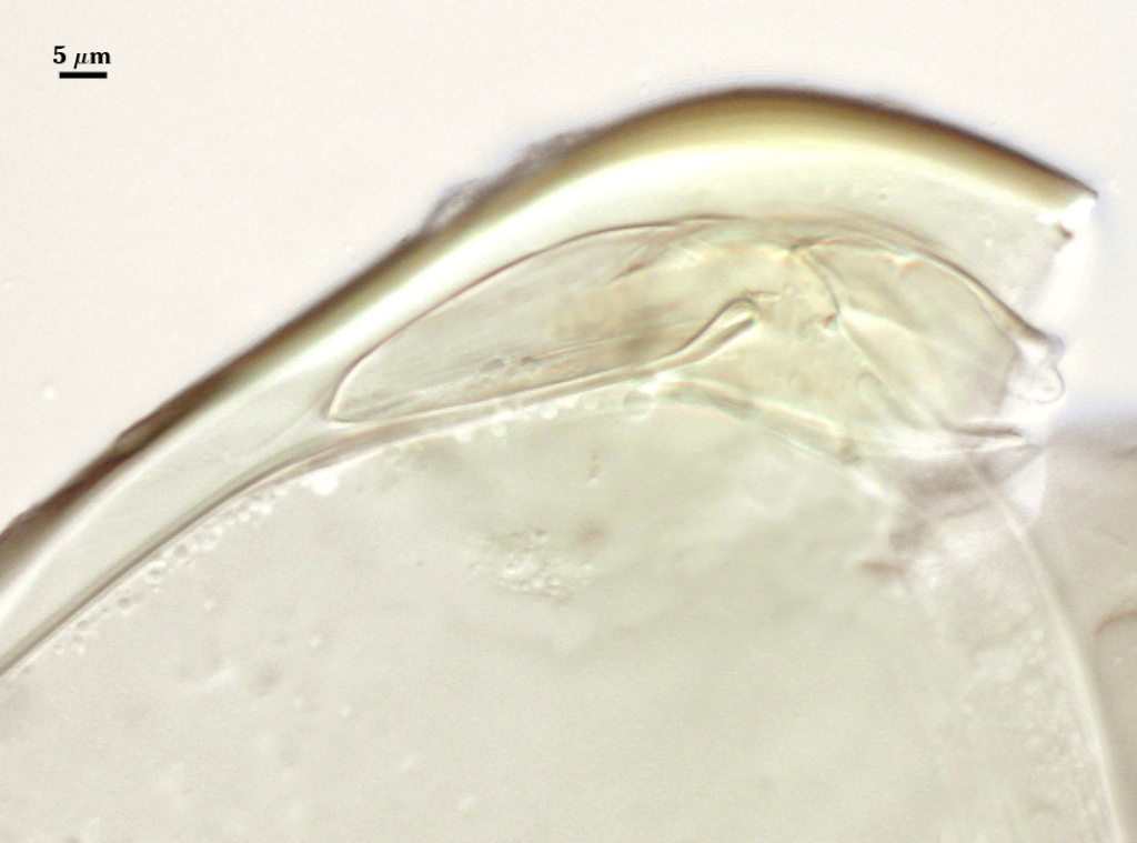
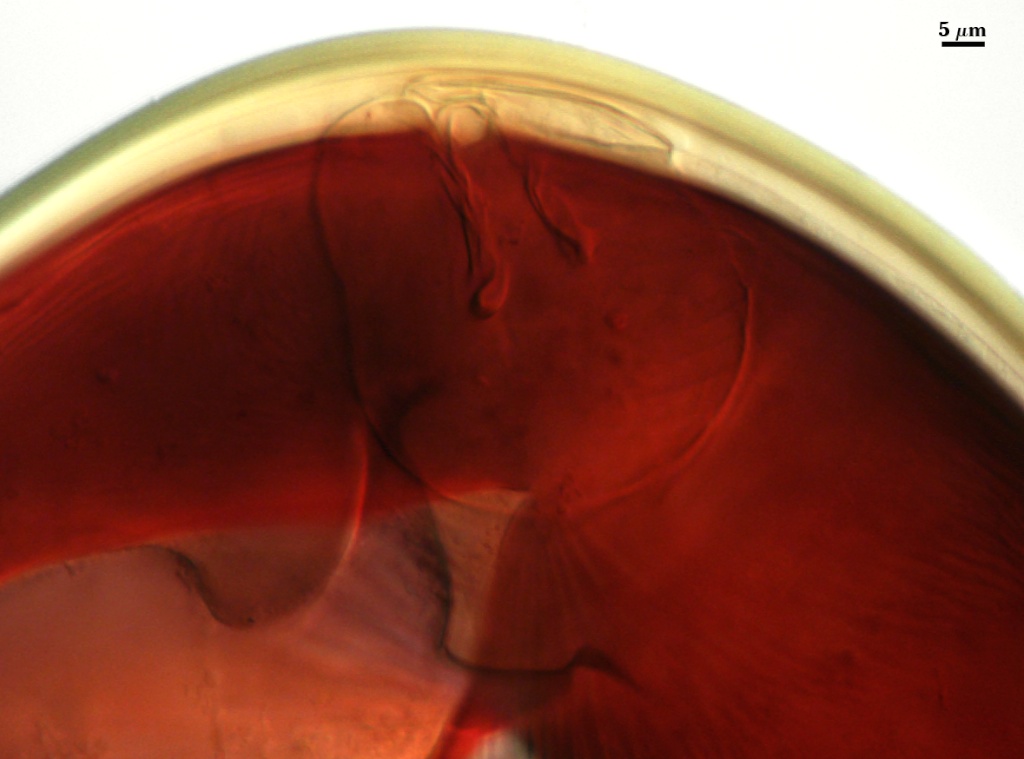
Subcellular Structure of Spores
During spore development, one spore wall consisting of two layers originated from the sporogenous cell wall are first differentiated, followed by independent differentiation of two bi-layered hyaline flexible inner walls (developmental sequence in photos below, from left to right).
| SPORE WALLS | ||||
|---|---|---|---|---|
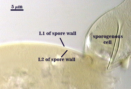 | 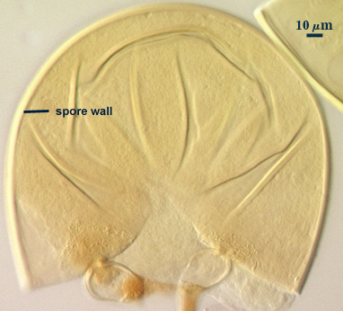 |  |  |  |
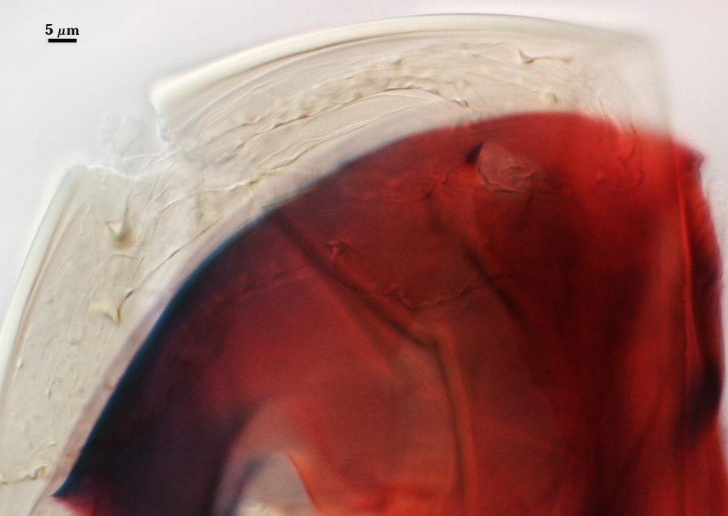
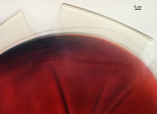
SPORE WALL: Two layers (L1 and L2) that are adherent that in juvenile spores are of equal thickness, with the laminate layer thickening as the spore wall is differentiated.
L1: An outer permanent rigid layer, smooth, pale yellow with a green tint (5-0-30-0), less than 1.0-1.2 µm thick and so tightly adherent to L2 that sometimes it is detectable only with superior optics under oil.
L2: A layer consisting of very fine adherent sublayers (or laminae) that together are 1.8-4.2 m thick (mean of 2.6 µm) in mature spores; pale yellow with a green tint (5-0-30-0). The innermost sublayers separate slightly and produce undulations that can be mistaken for an inner flexible wall.
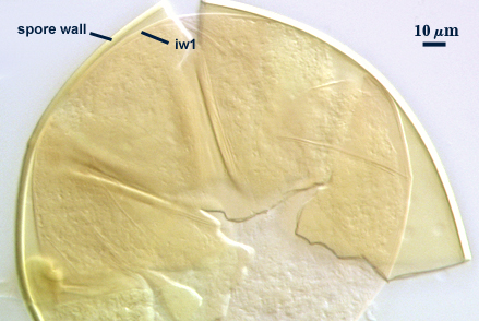
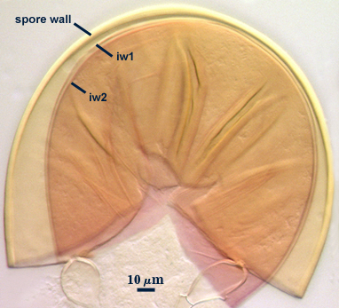
GERMINAL WALLS: Two hyaline flexible inner walls (gw1 and gw2) are formed, each with two adherent layers.
| GERMINAL WALLS | |||
|---|---|---|---|
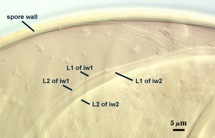 | 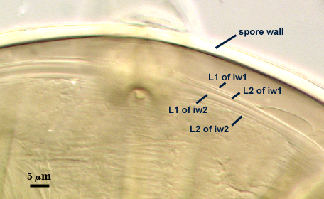 | 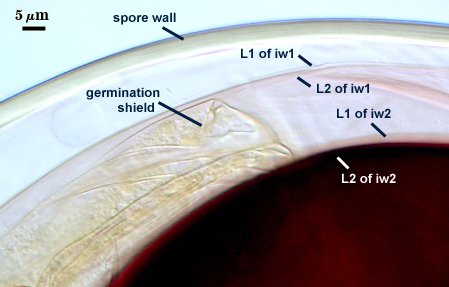 | 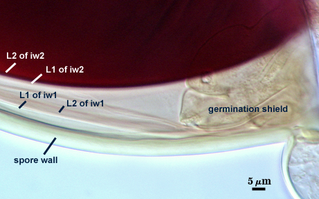 |
GW1: Two layers (L1 and L2) that are usually adherent and together are 0.9-2.0 µm thick. L1 is less than 0.5 µm thick; L2 is 0.5-1.4 µm thick. This wall often is positioned very close to the spore wall, and thus is difficult to detect amongst the undulations of sublayers in L2 of the spore wall(see above).
GW2: Two layers (L1 and L2) that almost always are adherent. L1 is 1.2-3.2 µm thick and often produces a weak pink reaction (0-10-20-0) in Melzer’s reagent that is detected only when it separates from the spore wall. L2 is hyaline and plastic enough that it has been termed “amorphous”. It is 2.5-8.0 µm thick in PVLG-based mountants, depending on amount of pressure applied to it while breaking the spore; staining red-purple (20-80-20-0) to dark red-purple (40-80-60-0) in Melzer’s reagent. This wall also does not separate much from the spore wall, even when considerable pressure is applied while breaking the spore, so that resolving iw2 from iw1 takes practice and examination of many spores (see photos of spores in PVLG above).
Subtending Hypha
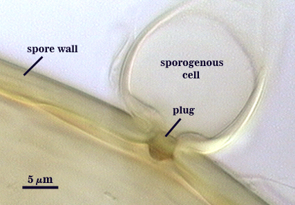
WIDTH OF SPOROGENOUS CELL: 22-28 µm (mean = 23.8 µm).
SPOROGENOUS CELL WALL: Two hyaline layers (L1 and L2) probably are present (continuous with the two layers of the spore wall), but only L2 is readily discernible at the level of the compound microscope.
L2: Pale yellow (0-0-20-0), 2.2-4.6 µm thick near the spore and then thinning to 0.6-1.0 µm beyond the sporogenous cell.
OCCLUSION: Closure by a plug concolorous with the laminate layer of the spore wall.
Germination
COLOR: Hyaline to pale yellow (0-0-20-0).
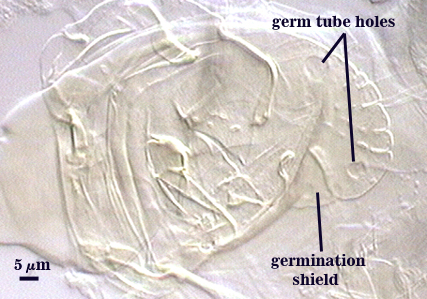
SHAPE: Ovoid to oblong, with length approximately 1.5 times that of the width. Margins of shields generally are smooth, with few folds (each with paired germ holes). The shield is fragile enough that it often folds or is easily fragmented. It also does not contrast from the inner walls with which it is associated and thus may be hard to detect. Position of the shield is on iw2.
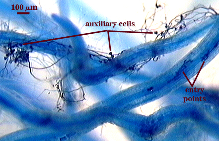
Auxiliary Cells
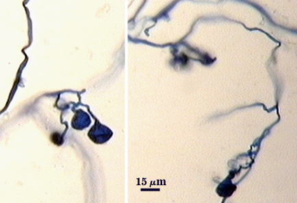
Cells often formed individually on closely spaced branches of coiled hyaline hyphae 3-4 µm in diameter; each cell thin-walled (< 1 µm thick), pale yellow (0-0-5-0) in transmitted light, each cell appearing almost smooth on the surface or with shallow swellings 0.5-1 µm high and 3-10 µm wide.
Mycorrhizae
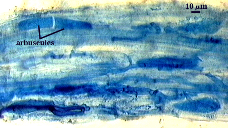
Intraradical arbuscules and hyphae consistently stain darkly in roots treated with trypan blue. Arbuscules with many fine tips from a swollen trunk. Hyphae often with knobs or projections, usually densely coiled near entry points.
| Arbuscules in corn | |
|---|---|
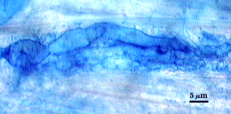 | |
| Other mycorrhizal structures in corn | ||
|---|---|---|
 |  | 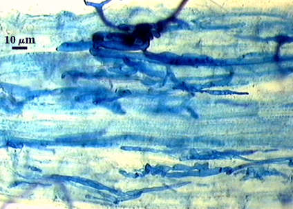 |
Notes
Spores resemble those of aged spores of S. pellucida under a dissecting microscope, except they have a smaller size range and few spores are oblong. They also are almost indistinguishable from spores of S. dipurpurascens, which is very similar in size, shape, and color and differs in its inner wall structure by having only one thin layer in gw1 instead of two layers.
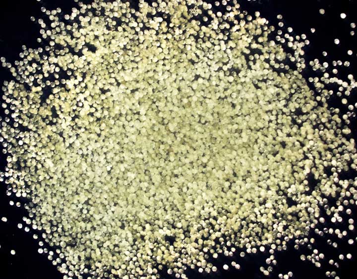
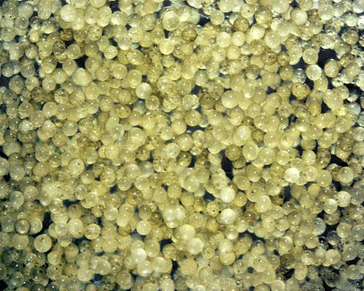
The images below can be uploaded into your browser by clicking on the thumbnail or can be downloaded to your computer by clicking on the link below each image. Please do not use these images for other than personal use without expressed permission from INVAM.
High Resolution Images | |
|---|---|
 |  |
 |  |
 |  |