Scutellospora dipurpurascens
(reference accession WV932)
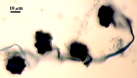
Whole Spores
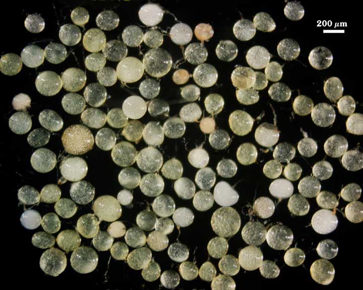
COLOR: Pale yellow with a greenish tint (5-0-20-0) to yellow-brown with greenish tint (5-0-100-10) in older spores.
SHAPE: Subglobose to oblong, sometimes irregular.
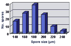
SIZE DISTRIBUTION: 140-240 µm, mean = 176 µm (n = 115).
Subcellular Structure of Spores
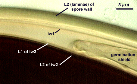
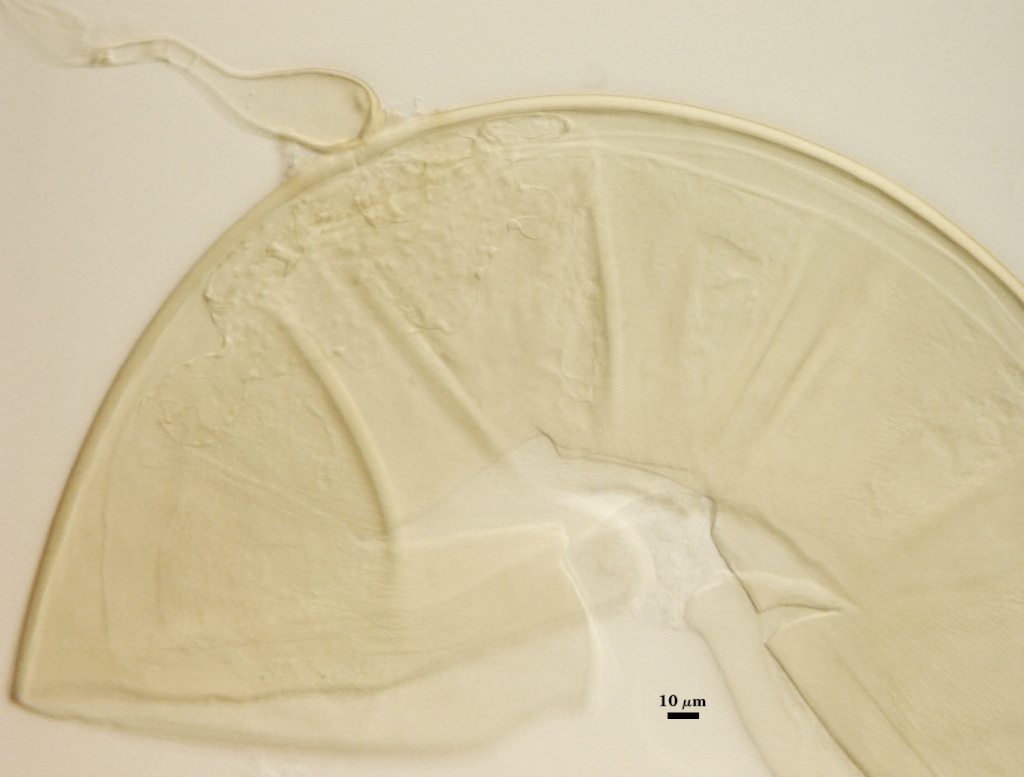
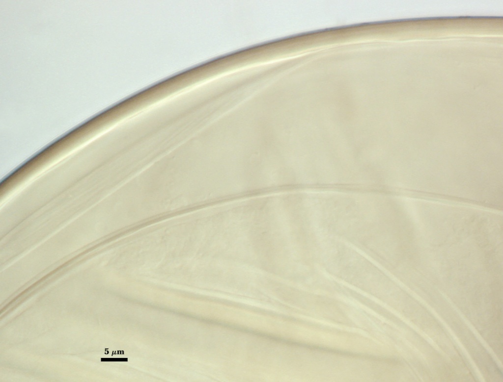
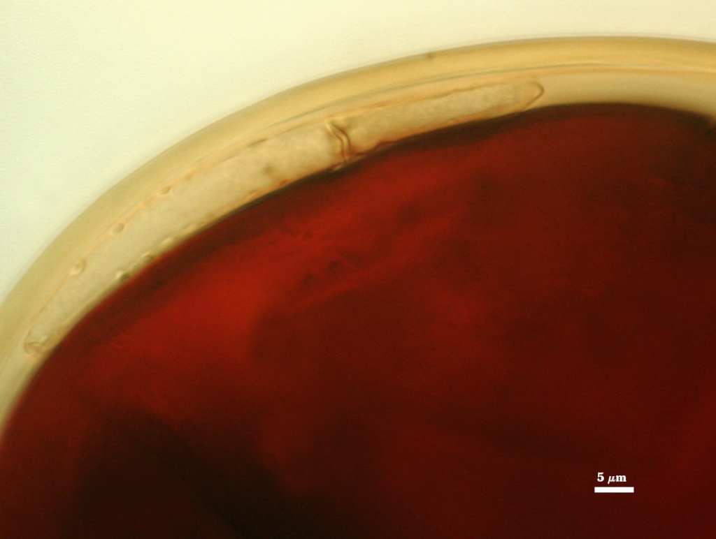
SPORE WALL: Two layers (L1 and L2) that are adherent that in juvenile spores are of equal thickness, with the laminate layer thickening as the spore wall is differentiated.
| In PVLG and Melzer’s reagent (1:1 v/v) | |||
|---|---|---|---|
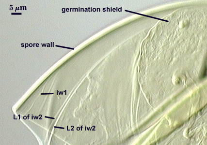 | 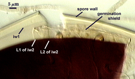 | 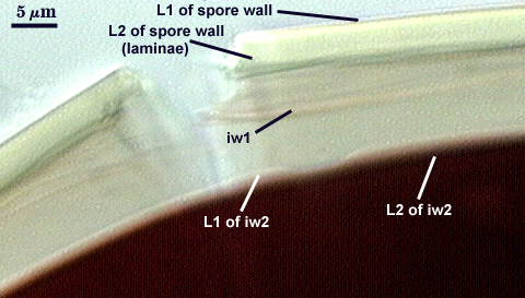 |  |
L1: An outer permanent rigid layer, smooth, pale yellow with a green tint (5-0-30-0), less than 1.0-1.2 µm thick and so tightly adherent to L2 that sometimes it is detectable only with superior optics under oil.
L2: A laminate layer consisting of very fine adherent sublayers (= laminations) that together are 1.8-4.2 m thick (mean of 2.6 m) in mature spores; pale yellow with a green tint (5-0-30-0). The innermost sublayers separate slightly and produce undulations that can be mistaken for an inner flexible wall.
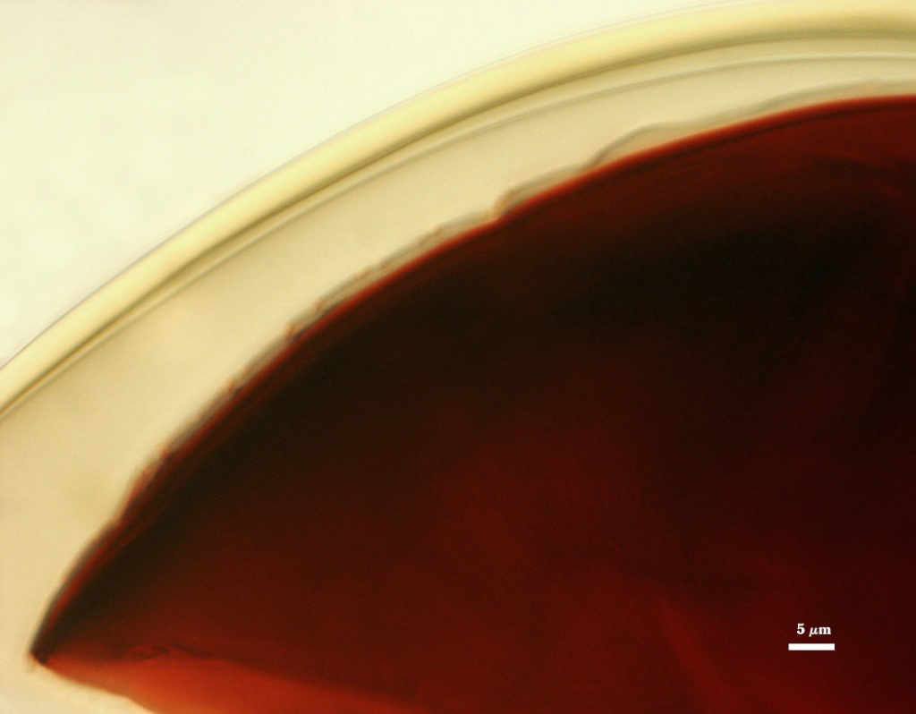
GERMINAL WALLS: Two flexible hyaline inner walls (gw1 and gw2) are formed.
GW1: One hyaline layer, 0.6-1.0 µm thick. It often is adherent to L2 (laminae) of the spore wall, at which time it is difficult to resolve (even under oil). It stains a light pink (0-20-20-0) in Melzer’s reagent, which is faintly observable when the layer separates distinctly from the spore wall (see photos above).
GW2: Two hyaline layers (L1 and L2) that almost always are adherent. L1 is 1.2-3.2 µm thick and often produces a weak pink reaction (0-10-20-0) in Melzer’s reagent that is detected only when it breaks away from the spore wall. L2 is hyaline and exhibits enough plasticity in acidic mountants to have been described as amorphous. It varies from 2.5-8.0 µm thick in PVLG-based mountants, depending on the degree of pressure applied to it while breaking the spore; stains red-purple (20-80-20-0) to dark red-purple (40-80-60-0) in Melzer’s reagent.
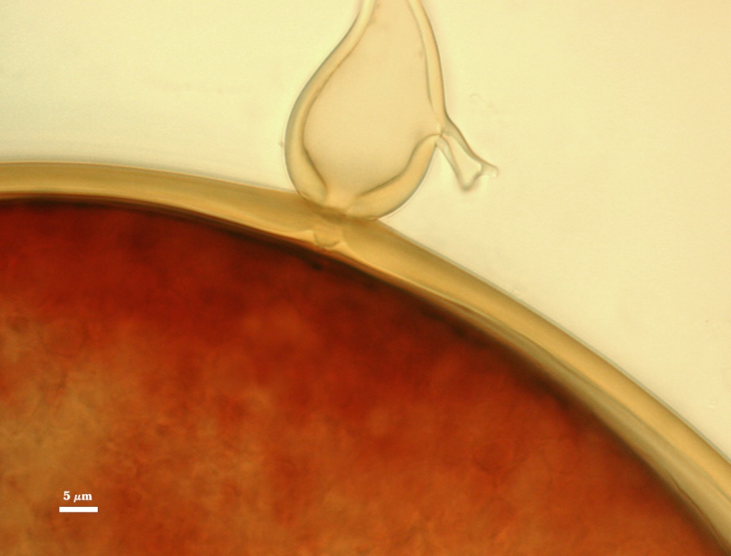
Subtending Hypha
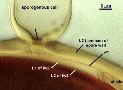
WIDTH OF SPOROGENOUS CELL: 21-30 µm (mean = 26.6 µm).
SPOROGENOUS CELL WALL: Two hyaline layers (L1 and L2) probably are present (continuous with the two layers of the spore wall), but only L2 is readily discernible at the level of the compound microscope.
L2: Pale greenish-yellow (5-0-20-0), 2.2-4.2 µm thick near the spore and then thinning to less than 1 µm beyond the sporogenous cell.
OCCLUSION: Closure by a plug concolorous with L2 of the spore wall.
Germination
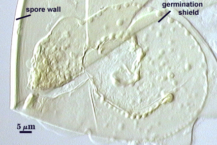
COLOR: Hyaline to pale greenish-yellow (5-0-20-0) because it is observed through pigments in the spore wall.
SHAPE: Oblong, with length approximately 1.5 times that of the width. Margins of shields generally are smooth, with few folds (most with paired germ holes). The shield is fragile enough that it often folds or is easily fragmented. It also does not contrast from the inner walls with which it is associated and thus may be hard to detect. Position of the shield is on iw2.
Auxiliary Cells
Aggregates of cells (1-10) borne on coiled hyaline to pale yellow (0-0-10-0) hyphae, thin-walled (< 1 µm thick), pale yellow (0-0-5-0) in transmitted light, each cell subglobose with almost smooth or with shallow swellings 0.5-2 µm high and 3-10 µm wide on the surface.
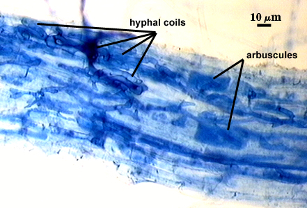
Mycorrhizae
Intraradical arbuscules and hyphae consistently stain darkly in roots treated with trypan blue. Arbuscules with many fine tips from a swollen trunk. Hyphae often with knobs or projections, usually densely coiled near entry points.
| Arbuscules in corn roots | Mycorrhizal structures in corn | ||
|---|---|---|---|
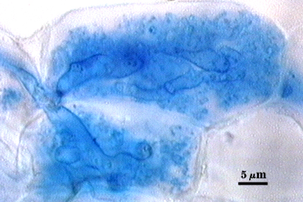 | 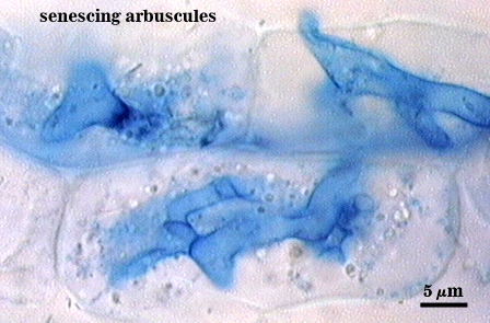 | 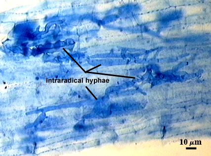 |  |
Notes
Spores resemble those of old spores of S. pellucida under a dissecting microscope, except they have a smaller size range and few spores are oblong. They also are almost indistinguishable from spores of S. calospora, which is very similar in size, shape, and color and differs in its inner wall structure except that it has two layers in gw1 rather than just one thin layer.
The images below can be uploaded into your browser by clicking on the thumbnail or can be downloaded to your computer by clicking on the link below each image. Please do not use these images for other than personal use without expressed permission from INVAM.
High Resolution Images | |
|---|---|
|  |
 |  |
 |  |