Acaulospora dilatata
(reference accession WV204)
Whole Spores
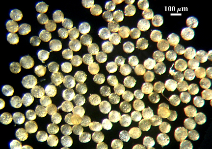
 COLOR: Pale yellow-brown (0-5-40-0) to pale yellow-brown (0-10-60-0)
SHAPE: Mostly globose to subglobose
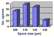
SIZE DISTRIBUTION: 100-130 µm, mean = 113 µm (n = 110))
Subcellular Structure of Spores
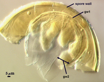
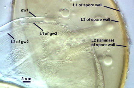
SPORE WALL: Three layers (L1, L2, and L3), the outer continous with the wall of the neck of the parent sporiferous saccule and the latter being synthesized with development of the spore.
L1: Hyaline, thin (< 0.5 µm), and sloughing with detachment of the saccule from the spore; rarely seen on detached spores.
L2: A layer consisting of very fine and adherent sublayers (or laminae), pale yellow-brown (0-10-60-0), 2.8-5.5 µm thick (mean = 4.7 µm). The layer surface is smooth once the outer layer has sloughed, which usually occurs (especially after sucrose-density centrifugation and washing). At maturity, the pore between spore and saccule neck is closed by continuous sublayers of this layer, sealing in spore contents.
L3: Pale yellow-brown (0-5-40-0), thin (1- 4 µm) and somewhat flexible when it separates from L2 of the spore wall. This layer also appears to consist of sublayers (laminae) which can separate more readily from each other than those of L2, so that they create folds that resemble numerous separate “inner walls”. In almost all instances, however, these sublayers merge as part of the spore wall. This layer is considered a discrete structure because it appears to be homologous with a layer of similar structure and position in other species (e.g., A. koskei, A. laevis). It has been described as “semi-rigid” by Schenck and coworkers because it breaks together with the spore wall (like gw1 described below).
| In PVLG | |
|---|---|
|
|
| In PVLG & Melzer’s reagent | ||
|---|---|---|
|
|
|
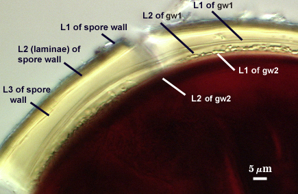
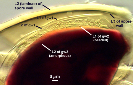
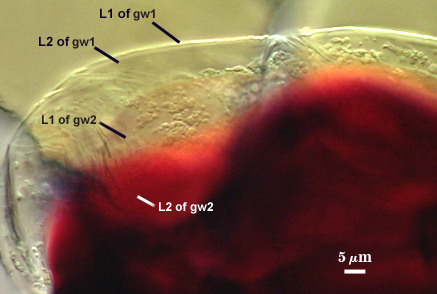
GERMINAL WALLS: Two flexible hyaline inner walls (gw1 and gw2) are present that readily separate from each other and from the spore wall.
GW1: A bilayered hyaline wall that separates easily from the spore wall and thus can be seen readily. Layers separate slightly in many spores, where they can be measured, but they also can be adherent and appear as a single layer. L1 is less than 0.5-1.0 µm thick, folding slightly when separated from L2. L2 is 0.5-1 µm thick. This wall has some inherent rigidity because it breaks with the spore wall.
GW2: Consisting of wo adherent hyaline layers. L1 is 0.5-1.2 µm thick (measurable only in PVLG), with granular excresences (or “beads”) that tend to become dislodged and float away with applied pressure. These “beads” are stabilized after preservation in formalin, but otherwise may be absent on mounted spores within a few months of storage. L2 is plastic enough that it has been termed “amorphous” (originally defined as an “amorphous wall”). It is <1.0 200 -7.0 µm thick in pvlg-based mountants, depending on amount of pressure applied to it while breaking the spore; staining red-purple (20-80-20-0) dark (40-80-60-0) melzer's reagent. Spores with more than once, l2 layer can become distended as far from spore.
Cicatrix
Scar indicating region of contact between spore and saccule neck during spore synthesis; very narrow lip (not measured) circumscribing the scar, circular to oval-shaped, 6-11 µm at widest (mean = 8.2 µm ).
Sporiferous Saccule
COLOR: Hyaline.
SHAPE: Mostly globose to subglobose, occasionally irregular.
SIZE: 100-135 µm, mean = 114 µm.
SACCULE WALL: One layer, smooth surface, 1.0-1.4 µm thick.
DISTANCE FROM SACCULE TO SPORE: 40-90 µm.
Germination
An ovoid “germination orb” forms on gw2, from which germ tubes form and penetrate through the spore wall. This orb is difficult to see except in older spores where contents have cleared with fusion of lipid globules in the spore lumen.
Notes
In older field collected spores, as well as spores stored for longer than 6 months in formalin or other preservatives, the beaded surface of gw2 becomes stabilized and the amorphous layer of gw2 becomes rigid (1.5-5 µm thick) and stains only faintly (light pinkish red or yell0w-brown) or not at all.
With a clearer picture of the morphological attributes of Acaulospora mellea, the comparison with A. dilatata is almost indistinguishable. The amorphous properties of the inner layer of gw2 are more pronounced in WV204 than in the reference isolate of A. mellea, but that is hardly a species-level distinction. It is probable that these two species are synonymous, and the only reason for delaying a taxonomic formalization of this change is the low number of isolates of both species available for comparative analysis.
The literature is replete with reports of A. mellea, but most of the fungi are likely A. morrowiae, which is almost identical morphologically except that it is smaller (usually < 80 µm) and lighter in color. Acaulospora morrowiae is a much more ubiquitous species and is by far the most common Acaulospora contaminant in greenhouse-grown pot cultures. It is a one of the most abundant sporulators in trap cultures from tropical soils.