Entrophospora infrequens
(reference accession AZ237)
Whole Spores | |
|---|---|
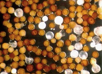 |  |
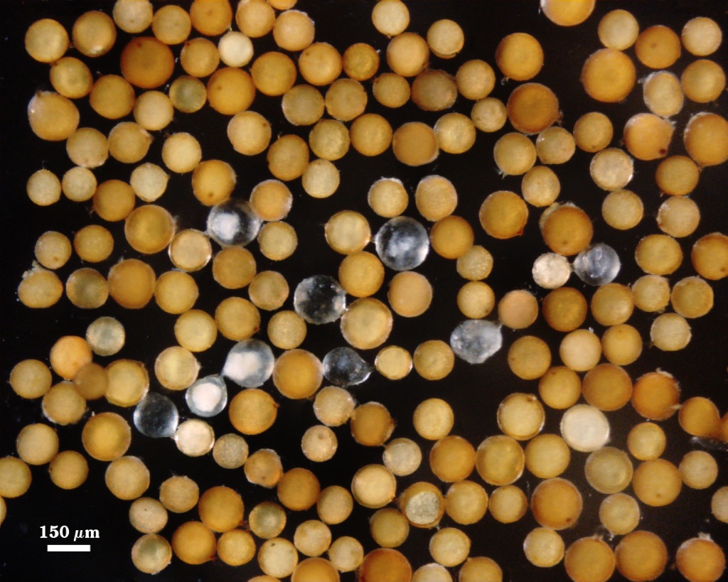
 COLOR: Light orange-brown (0-30-80-0) to dark orange-brown (0-60-100-0), most toward the darker orange.
SHAPE: Globose, some subglobose.
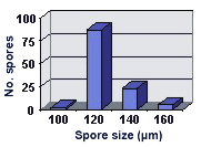
SIZE DISTRIBUTION: 100-160 µm, mean = 125.3 µm (n = 117).
Subcellular Structure of Spores
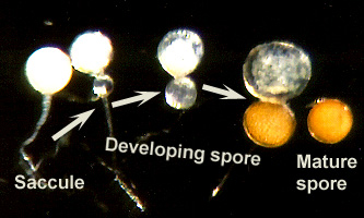
SPORE WALL: Four layers (L1, L2, L3 and L4), with L1-L3 continuous with the wall of the neck of the parent sporiferous saccule and L4 forming de novo. The appearance of whole spores under a stereomicroscope during this process can be seen in the first photo on the far left (A to D). All layers form sequentially during spore wall differentiation (other photos, from left to right).
| SPORE WALLS | ||||
|---|---|---|---|---|
 |  | 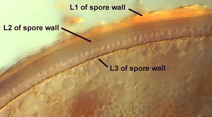 | 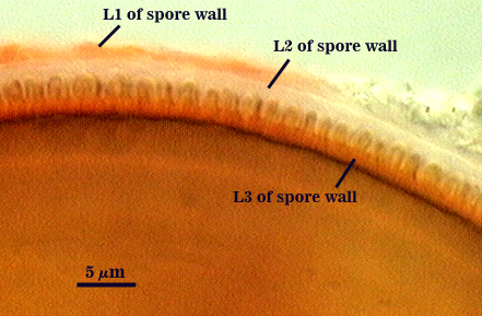 |  |
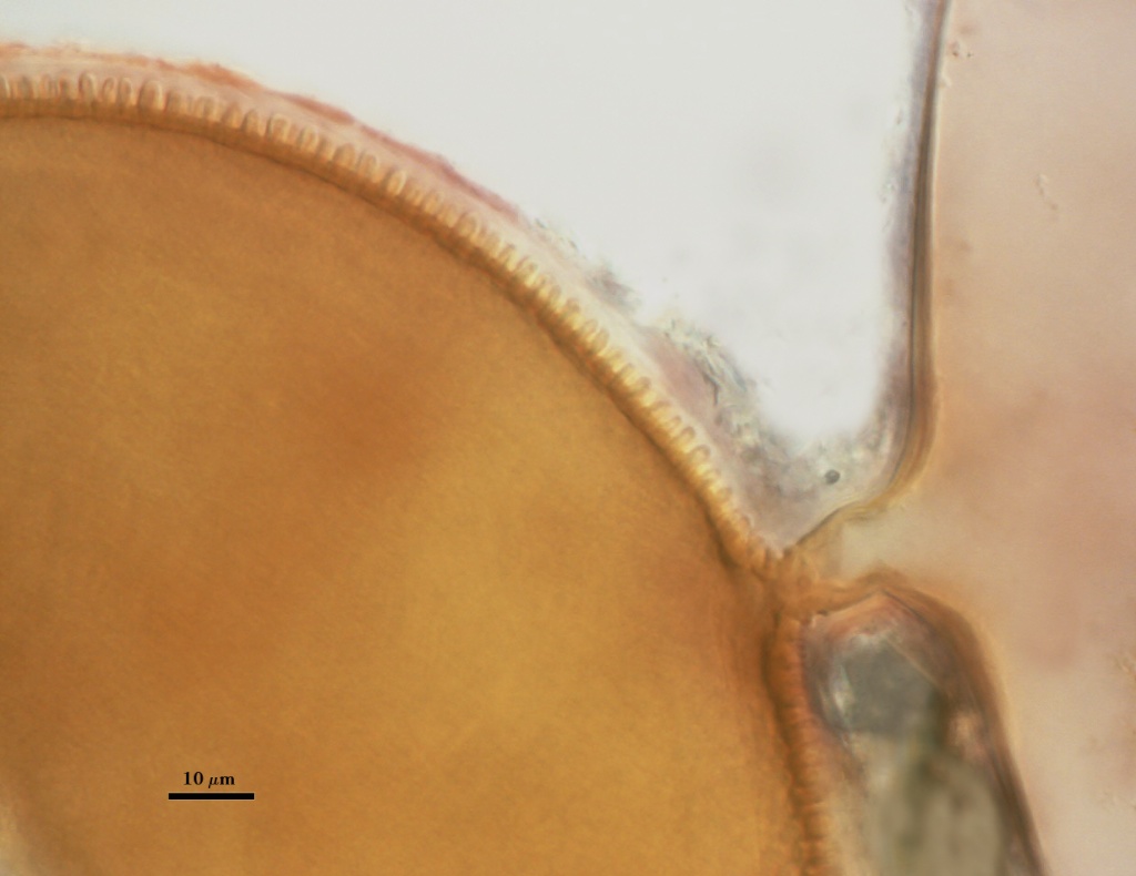
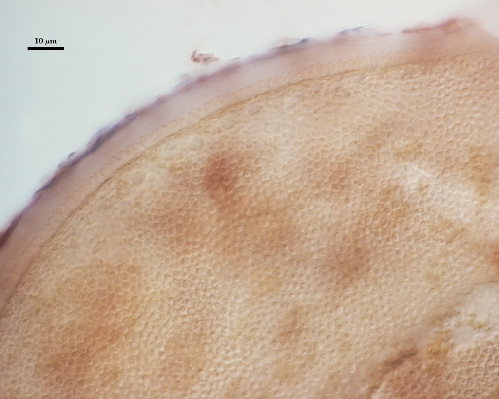
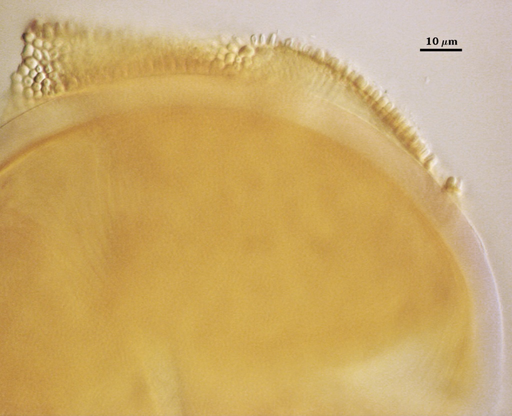
L1: The outermost hyaline layer is continuous with the outer layer of the saccule, degrading and sloughing as the spore ages; 2.0-5.0 µm thick when present intact (young spores), staining pink (0-20-20-0) to dark pinkish red (0-60-20-0).
L2: A hyaline layer, that initially is solid with shallow indentations on the inner surface and which later has imbedded in it the projections (or spines) of L3 (below) and then eventually degrades and sloughs; 3.5-5.0 thick (mean 4.4 µm) when present intact. This layer is very thin (< 0.5 µm) in the saccule wall and thus is difficult or impossible to see. Layer and spines together are 3.1-7.5 µm deep (6-7 µm in most spores).
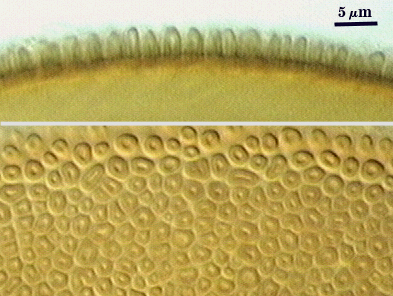
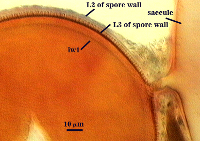
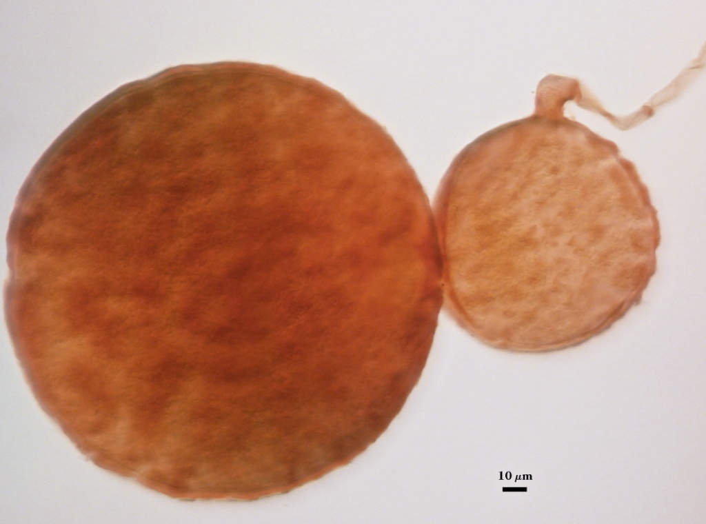
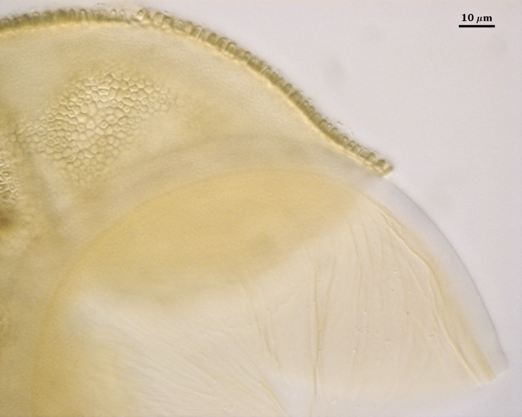
L3: A yellow (0-10-60-0) to yellow-brown (0-20-100-0) layer that initially is less than 0.5 µm thick and then expands to 1-1.4 µm thick just prior to formation of spines. No additional sublayers are formed as would be expected of a laminate layer (see A. delicata, A. morrowiae, etc.); instead, the width of this layer is determined by thickness of the basal layer plus height of spines. Each spine has five sides (pentagonal), 2.0-4.5 µm high, 1.3-3.0 µm wide (most 2.5 µm), and with a central depression 0.5-0.7 µm across. At maturity, the pore between spore and saccule neck is closed by a plug. The spore breaks away from the saccule neck at this point of attachment and appears sessile.
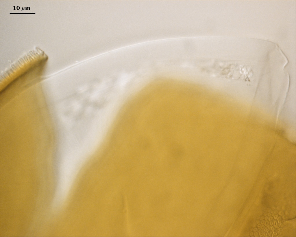
iw: Hyaline, semi-flexible, 7.5-9.2 µm thick (mean = 8.1 µm). This wall appears to be multi-layered, although the outer layer is so thin (< 0.3 µm) and adherent that it often cannot be detected. This component of the spore is very similar to the inner wall of species in Ambispora, suggesting this is a convergent trait.
Cicatrix
Two circular to ovoid scars indicating region of contact between spore and saccule neck during spore synthesis; it consists of tightly packed spines around a depression 15-30 µm (mean = 21.4 µm) wide on the cicatrix proximal to the saccule and 12-15 µm (mean = 13.8 µm) wide on the cicatrix distal to the saccule.
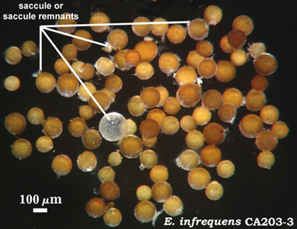
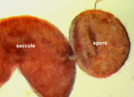
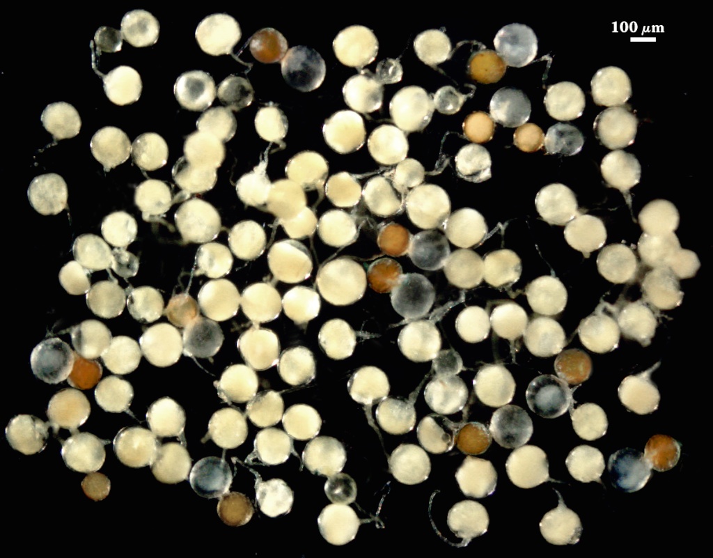
Sporiferous Saccule
COLOR: Hyaline.
SHAPE: Oblong.
SIZE DISTRIBUTION: 160-190 µm, mean = 174.5 µm.
SACCULE WALL THICKNESS: 3-11 µm.
SACCULE WALL: Three layers, 3-11 µm thick, continuous with layers of the spore wall. The outer layer is thickest in juvenile spores (5-19.8 µm) and is continuous with L1 of the spore. It appears to thicken from formation of numerous sublayers as well as have some mucilagenous properties (staining like L1 in Melzer’s reagent). The second layer (corresponding to L2 of the spore wall) is too thin to measure and often cannot be seen. The third layer (corresponding to L3 of the spore wall) is less than 0.3 µm except near the saccule hypha, where it thickens to 2.4 µm and is pale yellow (0-0-20-0).
DISTANCE FROM SACCULE TO SPORE: 12-15 µm.
Notes
In spores of intermediate age, spines from L3 of the spore wall are imbedded in the matrix of L2. However, L2 degrades over time, leaving the spines exposed.
The region between spore and saccule is unique among Entrophospora species in that the bridging hyphal wall has some pigmentation (pale yellow) and thickens appreciably near the spore. In addition, the pore between spore and saccule neck is closed by an amorphous plug rather than bridging layer(s) of the spore wall. The cicatrix distal to the saccule often is hard to detect in mature spores because it contains tubercles like the surrounding spore wall.
Cultures of this species have never seen sustained beyond one pot culture generation when the culture contents are stored away from living plants. This species has been maintained with other arbuscular fungi in a pot community and continues to sporulate as long as the pot culture is maintained (reseeding every 4-6 months).
The images below can be uploaded into your browser by clicking on the thumbnail or can be downloaded to your computer by clicking on the link below each image. Please do not use these images for other than personal use without expressed permission from INVAM.
High Resolution Images | |
|---|---|
 |  |
 |  |
 |  |
 |  |