Claroideoglomus luteum
(reference accession SA112)
| Accession | |
|---|---|
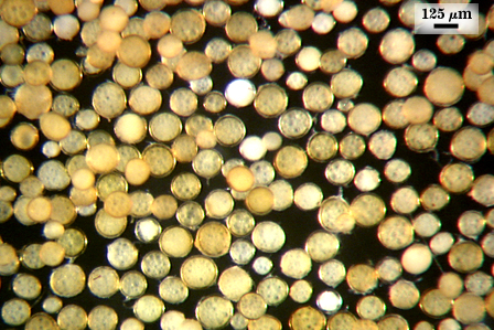 | 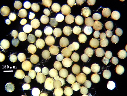 |
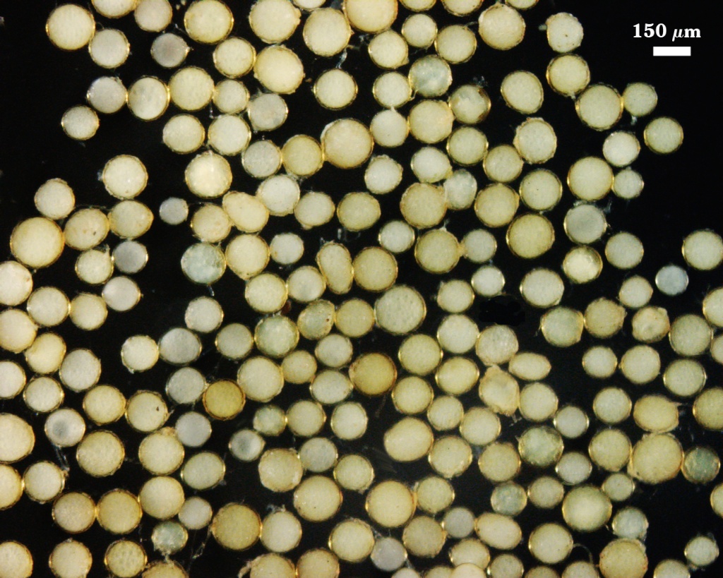
Whole Spores
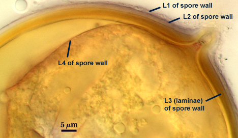
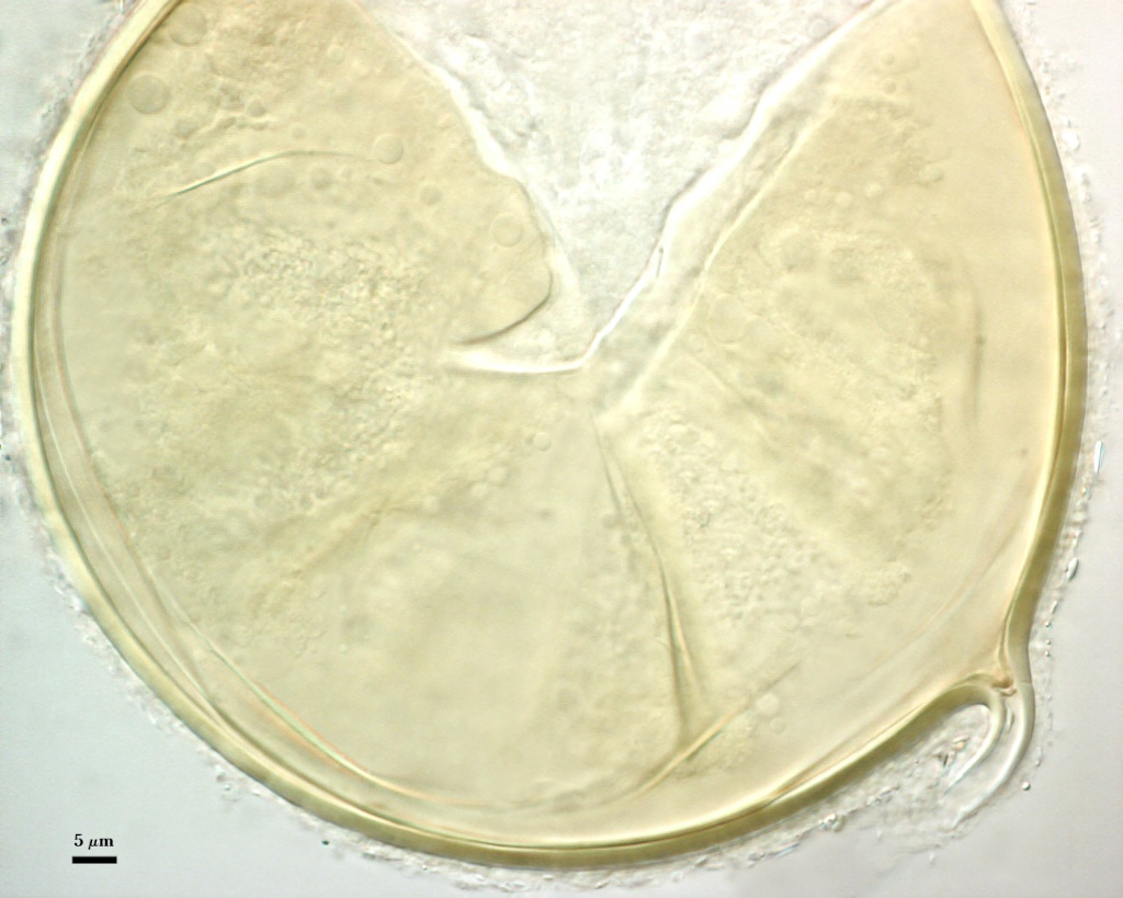
 COLOR: Pale yellow (0-0-20-0 to dark yellow with a brownish tint (0-10-60-0). Spores have a thin “halo” when all layers are present.
SHAPE: Globose to subglobose usually
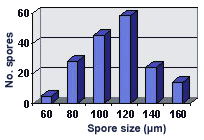
SIZE DISTRIBUTION: 60-180 µm, mean = 110 µm (n = 164)
Subcellular Structure of Spores
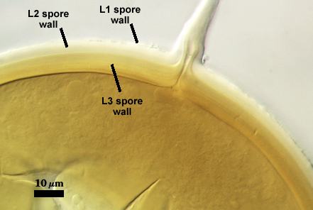
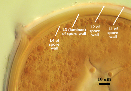
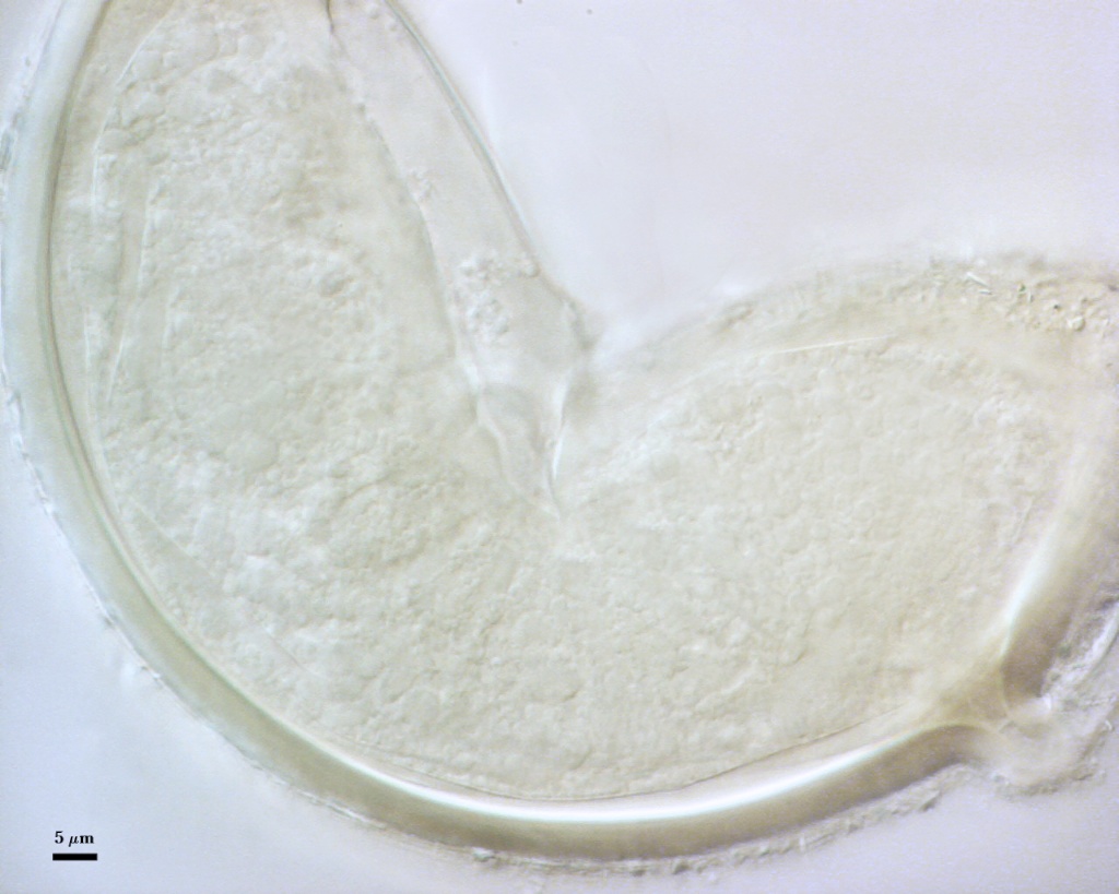
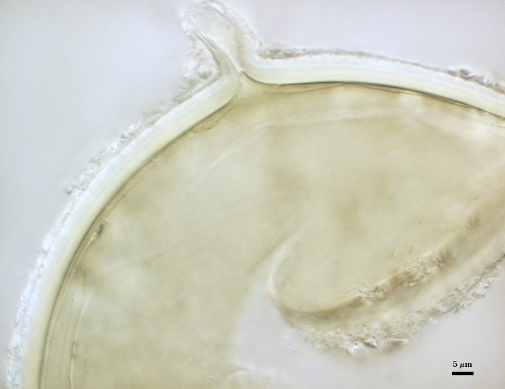
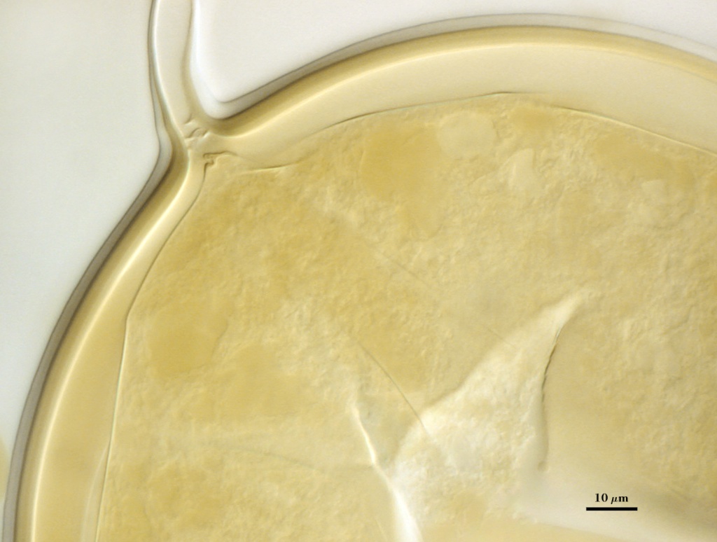
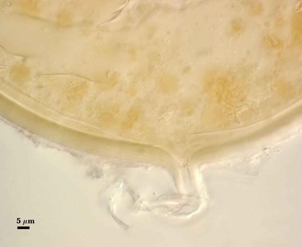
SPORE WALL: Four layers (L1, L2, L3 and L4), all are adherent, with L4 often separating in mature spores with applied pressure. These layers form consecutively as the spore wall differentiates (left to right in sequence of photos below), with only L1 and L2 present in the most juvenile spores.
| Luteum | ||||
|---|---|---|---|---|
|
|
|
|
|
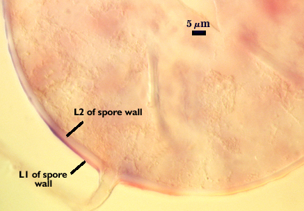
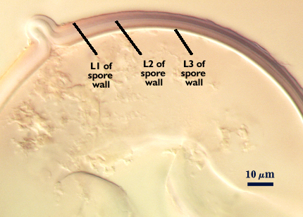
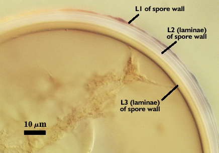
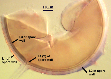
L1: Hyaline mucilagenous layer with considerable variation in thickness, 1.3-5.0 µm; intact on young spores and then degrading as the spore matures. In older spores, this layer may be completely absent.
L2: Hyaline, semi-rigid, 1.3-6.5 µm thick (mean = 2.5 µm), producing no reaction in Melzer’s reagent. This layer also forms with L1 on juvenile spores and begins to degrade as the spore matures. In pot culture, this layer is present on over 50% of mature spores, but with some degradation evident. On other spores, this layer is absent.
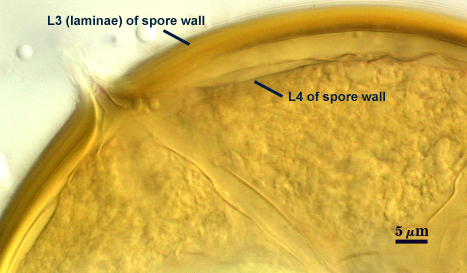
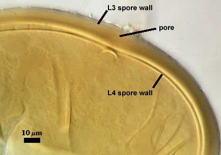
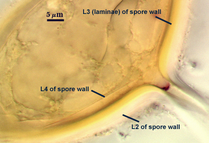
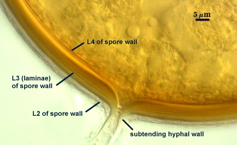
L3: Rigid permanent layer, consisting of fine adherent sublayers (or laminae), pale yellow (0-0-20-0) to brownish yellow (0-10-60-0), 2.5-10 µm thick (mean = 5.5 µm). This layer extends into the subtending hypha.
L4: A thin flexible layer concolorous with the laminate layer, very thin (< 0.5 µm), forming folds and wrinkles in broken spores. Its appearance closely resembles that of a germinal wall (= “membranous” wall), but its development and function are quite different: originating as part of the subtending hyphal wall and having no role in germination processes. It is considered a separate layer rather than a sublayer of L3 because it consistently separates to varying degrees from the spore wall in at least 75% of spores in populations of all accessions. When adherent to L3, it cannot be seen. In some spores it breaks away completely from the spore wall, and it has a small protrusion where it was attached to the subtending hyphal wall.
| In PVLG | |
|---|---|
|
|
| In PVLG and Melzer's Reagent (1:1 v/v) | ||
|---|---|---|
|
|
|
Subtending Hypha
SHAPE: Cylindrical to slightly flared (see photos above).
WIDTH: 9-19 µm
WALL STRUCTURE: The wall of the subtending hypha differentiates simultaneously with layers of the spore wall (see developmental sequence above), with the outer three layers extending the length of the hypha. The innermost layer (L4) only extends into the hypha lumen to the occlusion structure. The subtending hypha has a tendency to break off at the spore base and thus go undetected. When present, only the pigmented (yellow-brown) layer (L3) is permanent and the outer hyaline layer (L2) is present at varying thickness (depending on state of degradation).
OCCLUSION: Innermost sublayer of L3 of the spore wall occludes the hypha lumen, or a thin septum forms from L3.
Germination
A germ tube emerges from the lumen of the subtending hypha, and originates either at the septum or at a break in branch hyphae.
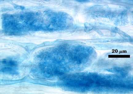
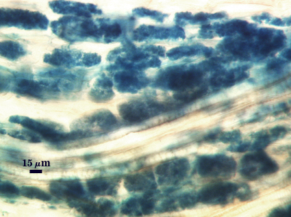
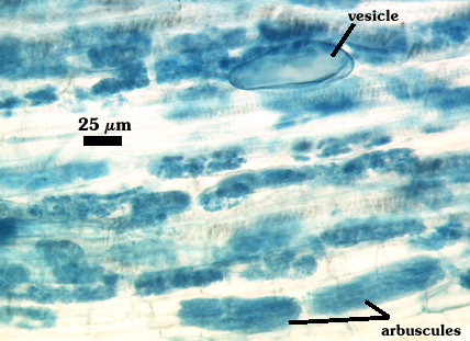
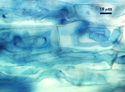
Mycorrhizae
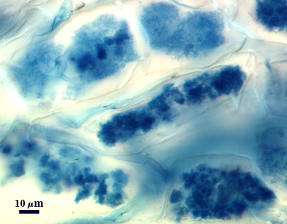
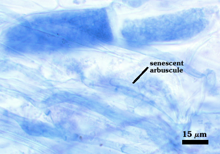
Fungal structures in mycorrhizae generally stain darkly, although lighter regions also are observed. Arbscules consist of trunks 1.5-4 µm wide with branch hyphae usually thinging incrementally to fine tips. Vesicles are rare, and then only sporadically distributed. When present, vesicles are ellipsoid, 32-59×75-95 µm diam. Hyphae at entry points often are coiled, 4-9 µm wide. Colonizing hyphae grow straight and parallel to the root axis, generally 2-5 µm wide. Infection units often overlap so that there are long, continuous regions of mycorrhizal colonization.
| Arbuscules in corn roots | ||
|---|---|---|
|
|
|
| All mycorrhizal structures in corn roots | ||
|---|---|---|
|
|
|
Notes
Spores of C. luteum are within the size and color range of many other glomoid species, but they most closely resemble those of R. clarus and C. claroideum. In fact, the holotype of this species is identical to another accession from the same region (SA101) that has been reported as G. clarum isolate NT4 (Talukdar and Germida 1993a, 1993b; Walley and Germida 1996). The distinctions in spore wall phenotype are summarized as follows:
L1: Hyaline and mucilaginous in all three species, but it is most darkly dextrinoid in C. clarus and lightest in G. claroideum.
L2: Hyaline, degrades from spores of C. claroideum and C. luteum, but is permanent and much thicker in R. clarus. Thickness and retention of this layer gives spores of C. luteum a halo appearance under reflected light (see photo at top of page), which is even more pronounced on spores of C. clarus.
L3: Pigmented (pale to dark yellow) laminate layer that is thickest in C. luteum, of intermediate thickness in C. claroideum, and thinnest in R. clarus. The greater thickness of this layer in C. luteum accounts for the darker yellow appearance of many spores in comparison to those of C. claroideum.
L4: Only present in spores of C. luteum and C. claroideum, with similar structure and position in the lumen of the subtending hypha.
The images below can be uploaded into your browser by clicking on the thumbnail or can be downloaded to your computer by clicking on the link below each image. Please do not use these images for other than personal use without expressed permission from INVAM.
High Resolution Images | |
|---|---|
 |  |
 |  |
 |  |
Reference
- Talukdar, N. C. and J. J. Germida. 1993a. Occurrence and isolation of vesicular-arbuscular mycorrhizae in cropped field soils of Saskatchewan, Canada. Can. J. Microbiol. 39:567-575.
- Talukdar, N. C. and J. J. Germida. 1993b. Propagation and storage of vesicular-arbuscular mycorrhizal fungi isolated from Saskatchewan agricultural soils. Can. J. Bot. 71:1328-1335.
- Walley, F. J. and J. J. Germida. 1996. Failure to decontaminate Glomus clarum NT4 spores is due to spore wall-associated bacteria. Mycorrhiza 6:43-49.