Glomus maculosum
Whole Spores
This species has been synonymized with Glomus claroideum (Walker and Vestberg, 1998), a move long overdue. Type specimens show strong evidence for structural modifications of spores as a result of parasitism. The description below is that of Miller and Walker (1986) with terminology modified to match that used throughout this web site.
Spores are formed singly in the soil on one to three subtending hyphae; hyaline when immature, becoming pale straw-colored to ochraceous when mature, globose to subglobose, (95-)135-178(-220) x (95-)130-187 (-220) µm.
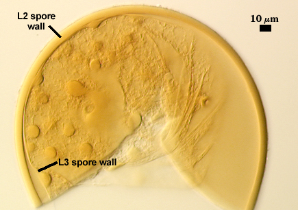
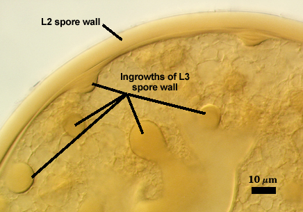
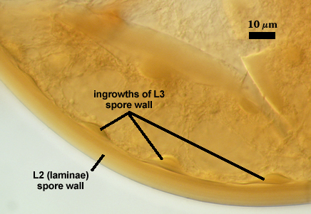
Spore wall structure of three layers (L1, L2 and L3). The outer layer (L1) is thin, hyaline, 0.3-1.0 µm thick, tightly adherent to L2, sloughing with age. The middle layer consists of finely adherent sublayers (or laminae), pale straw-colored to ochraceous, 4-13 µm thick. The innermost layer is 0.5-1 µm thick, flexible, and readily separating from L2 with age. In older spores, L3 forms domed, scalloped ingrowths, 6-15 µm wide and up to 12 µm deep, consisting of 2-8 concentric bulging discs increasing in diameter towards the inside of the spore.
Subtending hypha concolorous with L2 of the spore wall, straight to sharply recurved, parallel-sided, or funnelshaped, sometimes constricted at the spore base, 5-13(-25) µm long, 5-13 µm wide proximally and 5-7 µm wide at the point of connection to a thin-walled, hyaline parent hypha.
| Holotype specimens mounted in PVLG | ||
|---|---|---|
 |  |  |
Notes
Use of the term “membranous” to define the innermost layer of this species (and a number of other species in Glomus) and treat this structure as identical to those of similar appearance in other families (Acaulosporaceae, Gigasporaceae) creates considerable confusion because it implies that they are somehow comparable. Such an approach is erroneous phylogenetically, because this structure in Glomus has no similarities (not even as an analog) in other families. It clearly is part of the spore wall ontogenetically and has none of the involvement in pregermination processes seen in spores of other families. In fact, this layer can vary considerably in thickness and number of sublayers so that it also could be mistaken for a “laminate” layer (see range of phenotypes on the G. claroideum page). Similarly, the unusual “maculosa” erroneously thought to be a grouping criterion for this species also is found in parasitized spores of other species with a thin inner layer (see example on Glomus geosporum page).