Archaeospora schenckii
(reference accession CL401)
=Entrophospora schenckii
Whole Spores
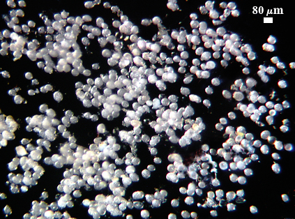
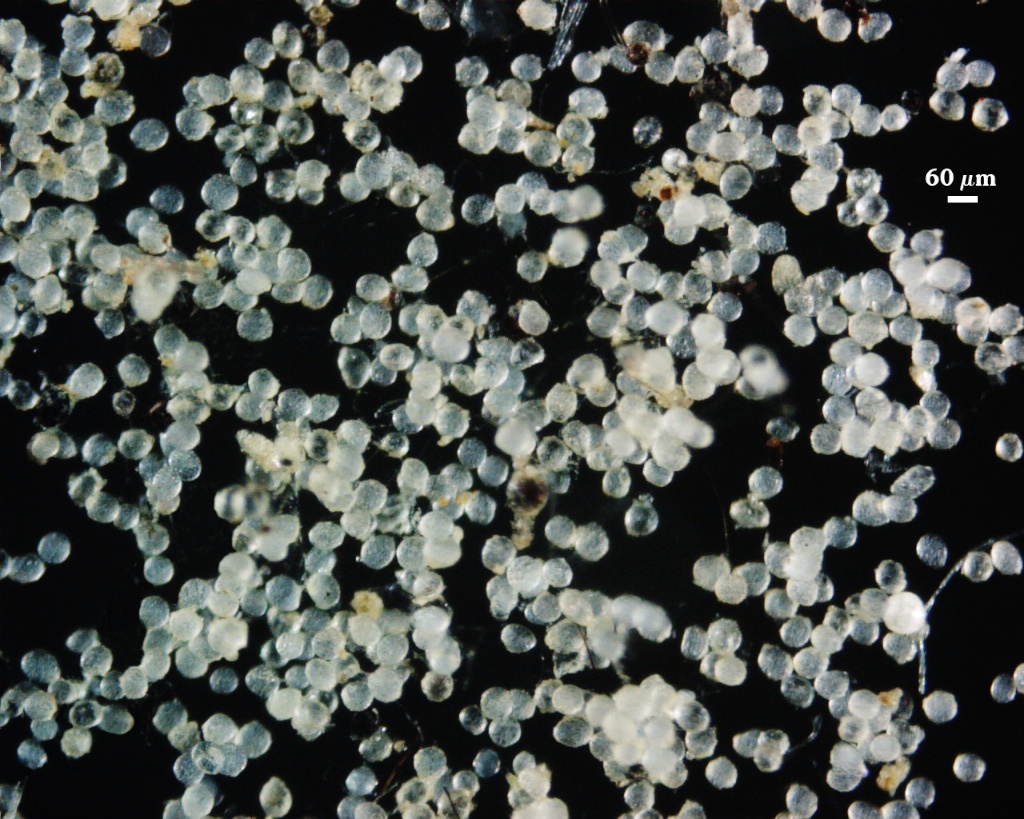
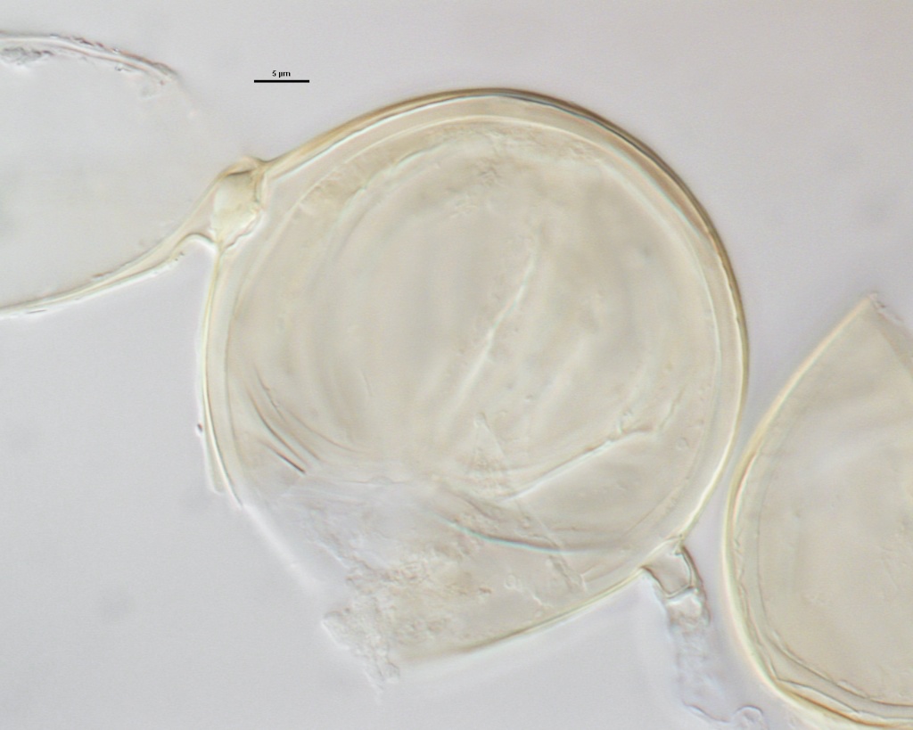
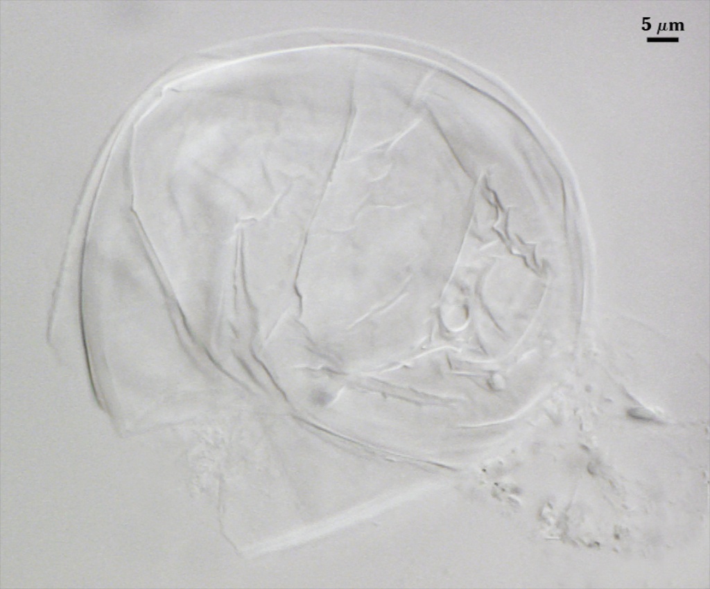
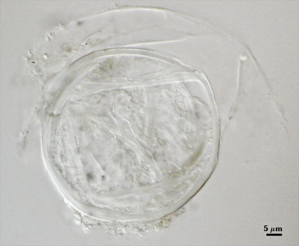
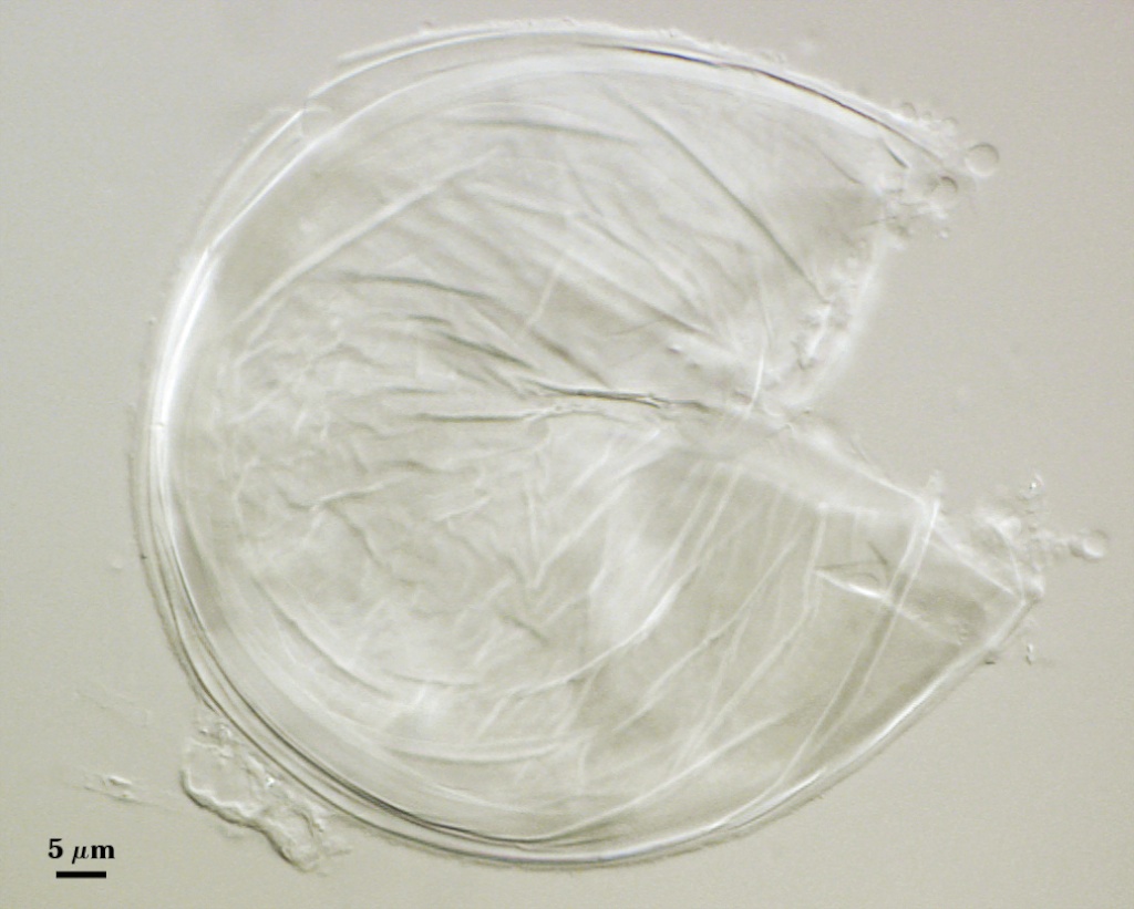
 COLOR: Completely hyaline (sparkling white) when mature.
SHAPE: Mostly globose, subglobose, but also ellipsoid to ovoid.
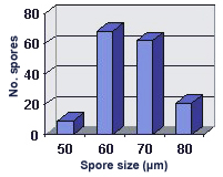
SIZE DISTRIBUTION: 50-80 µm, mean = 64 µm (n = 102)
Subcellular Structure of Spores
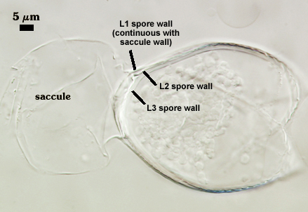
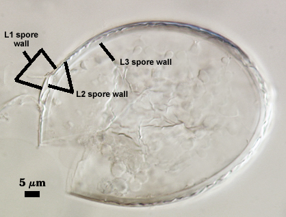
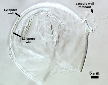
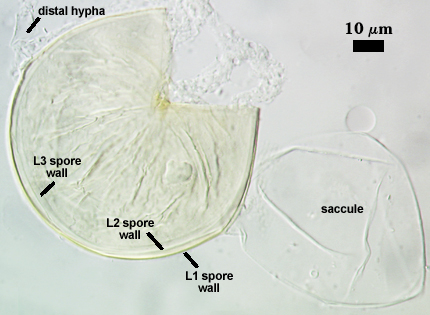
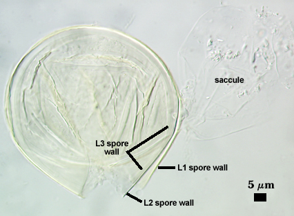
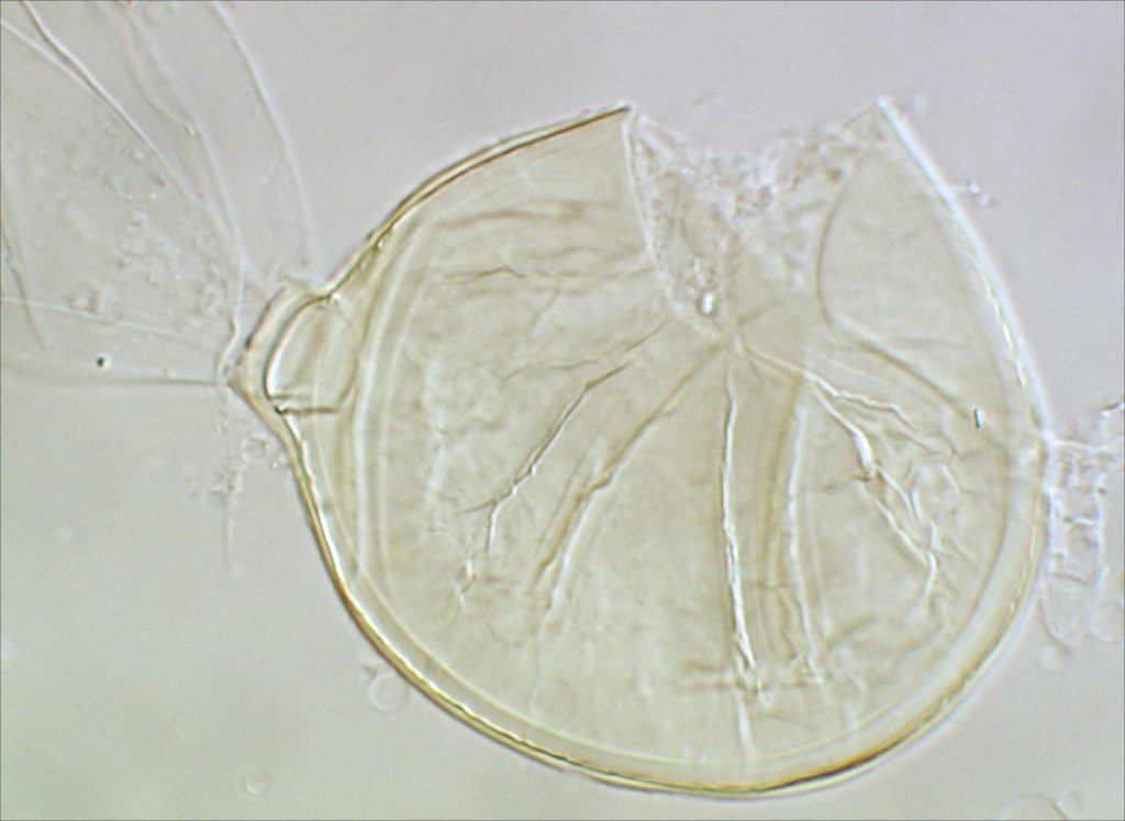
SPORE WALL: Consisting of three hyaline layers (L1, L2 and L3), all of which exhibit some flexibility in broken section. As a result, the spore wall contains numerous folds from any or all of the layers.
| In PVLG | |
|---|---|
 |  |
| In PVLG & Melzer’s reagent | |||
|---|---|---|---|
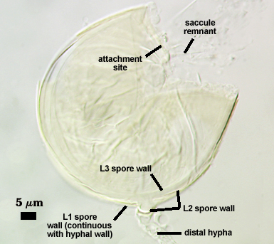 | 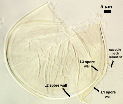 | 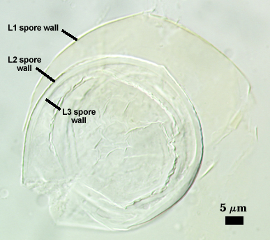 | 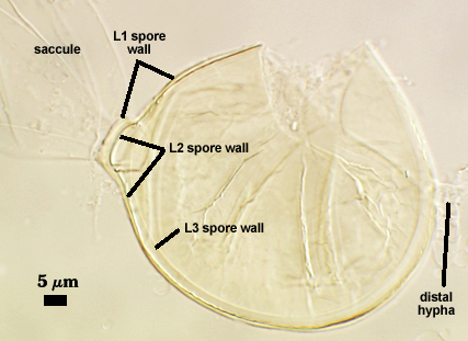 |
L1: A thin hyaline layer, < 1 µm thick, which is continuous with the wall of the neck of the sporiferous saccule. This layer often separates somewhat from the inner two layers and sometimes sloughs completely with loss of the saccule.
L2: A thin hyaline layer, < 1 µm thick, which usually is adherent to the innermost layer (L3). It has distinct margins that resolve it’s discrete structure; in some spores it may separately slightly from L3.
L3: A thicker hyaline layer than either L1 or L2, ranging from 1.3-4 µm thick, which together with L2 form an endospore which encloses the spore contents.
Cicatrix
Cicatrix proximal to saccule is 7-10 µm in diameter, with a very thin < 1 µm ridge. The smaller distal cicatrix rarely is seen to measure (usually attached hypha is absent and there is no defining detail by which to find it).
Sporiferous Saccule
COLOR: Hyaline.
SHAPE: Mostly subglobose, occasionally ellipsoid
SIZE DISTRIBUTION: 50-70 µm, mean = 59 µm.
SACCULE WALL: One layer, sometimes with an outer coating of granular material, < 0.5-1.0 µm.
DISTANCE FROM SACCULE TO SPORE: 10-30 µm.
| Schneckii | |
|---|---|
 |  |
Mycorrhizae
Light staining, much like that reported by Sieverding and Toro (1987) and likely a trait shared by Ambispora species. Given that pattern, this trait likely is a family level synapomorphy (uniquely defining the Archaeosporaceae clade.
Notes
When spores float in water after extraction, the saccule often is oriented downward so that it is not always easy to detect, much like that of Archaeospora trappei. When saccules are absent, spores are almost impossible to distinguish from Ar. trappei under a dissecting microscope. When remnants of the saccule neck are present (suggestive of a subtending hyphae), then spores are easily mistaken for Paraglomus occultum.
Archaeospora schenckii is unique from species in Acaulospora in that spore subcellular structure is unique. There are no flexible inner walls, hence germination must occur through the spore wall. Interestingly, this same subcellular structure is the phenotype of Ar. trappei spores. Clearly, they both belong in the same genus, which Schüßler and Walker (2010) formalized. Now the question arises: Are they two morphs of the same species? We have resolved that question and a publication is in preparation.
The images below can be uploaded into your browser by clicking on the thumbnail or can be downloaded to your computer by clicking on the link below each image. Please do not use these images for other than personal use without expressed permission from INVAM.
High Resolution Images | |
|---|---|
 |  |
 |  |
 |  |
Links to Gene Sequences in Genbank
Reference
Sieverding, E. and S. Toro. 1987. Entrophospora schenckii: A new species in the Endogonaceae from Colombia. Mycotaxon 28:209-214.
Schüßler, A and C. Walker C. 2010. The Glomeromycota: a species list with new families. Electronic copy available online at Glomeromycota PHYLOGENY.