Archaeospora trappei
(reference accession AU219)
= Acaulospora trappei
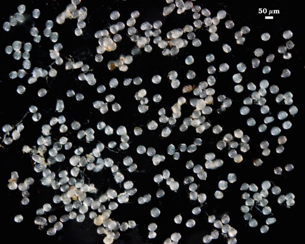
Whole Spores
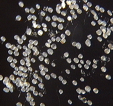
 COLOR: Usually completely hyaline (sparkling) and rarely creamy white when immature (0-0-10-0)
SHAPE: Mostly globose, subglobose, occasionally irregular.
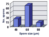
SIZE DISTRIBUTION: 40-80 µm, mean = 57.8 µm (n = 109)
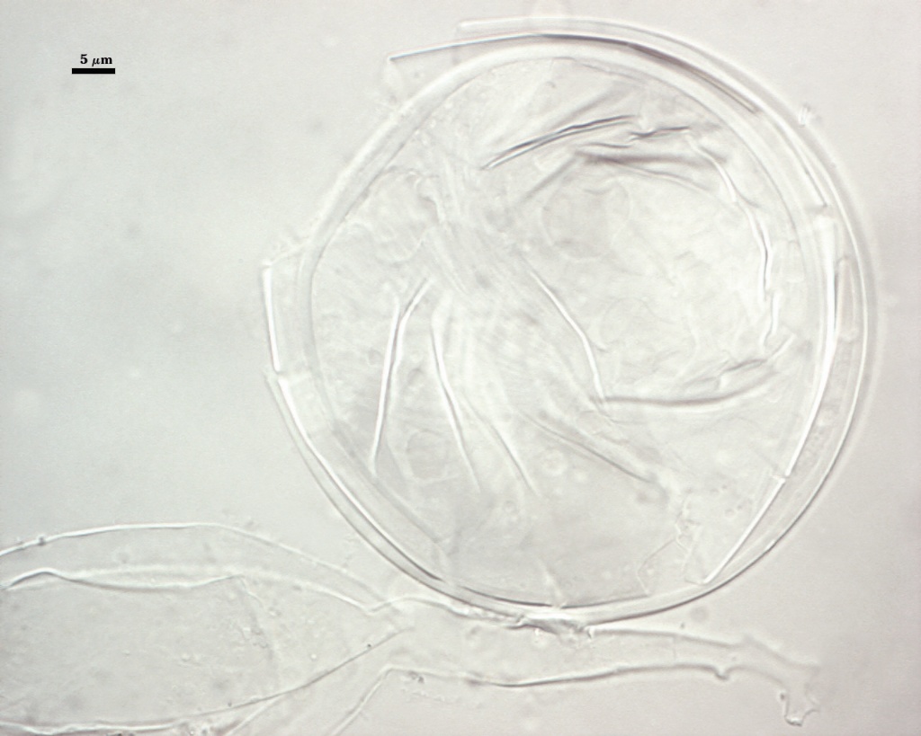
Subcellular Structure of Spores
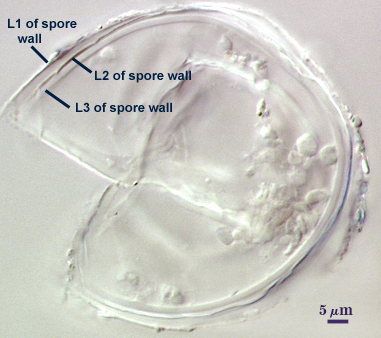
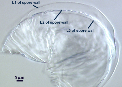
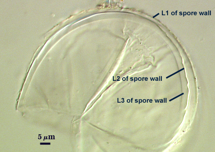
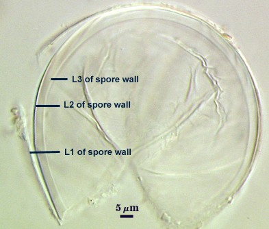
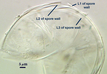
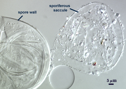
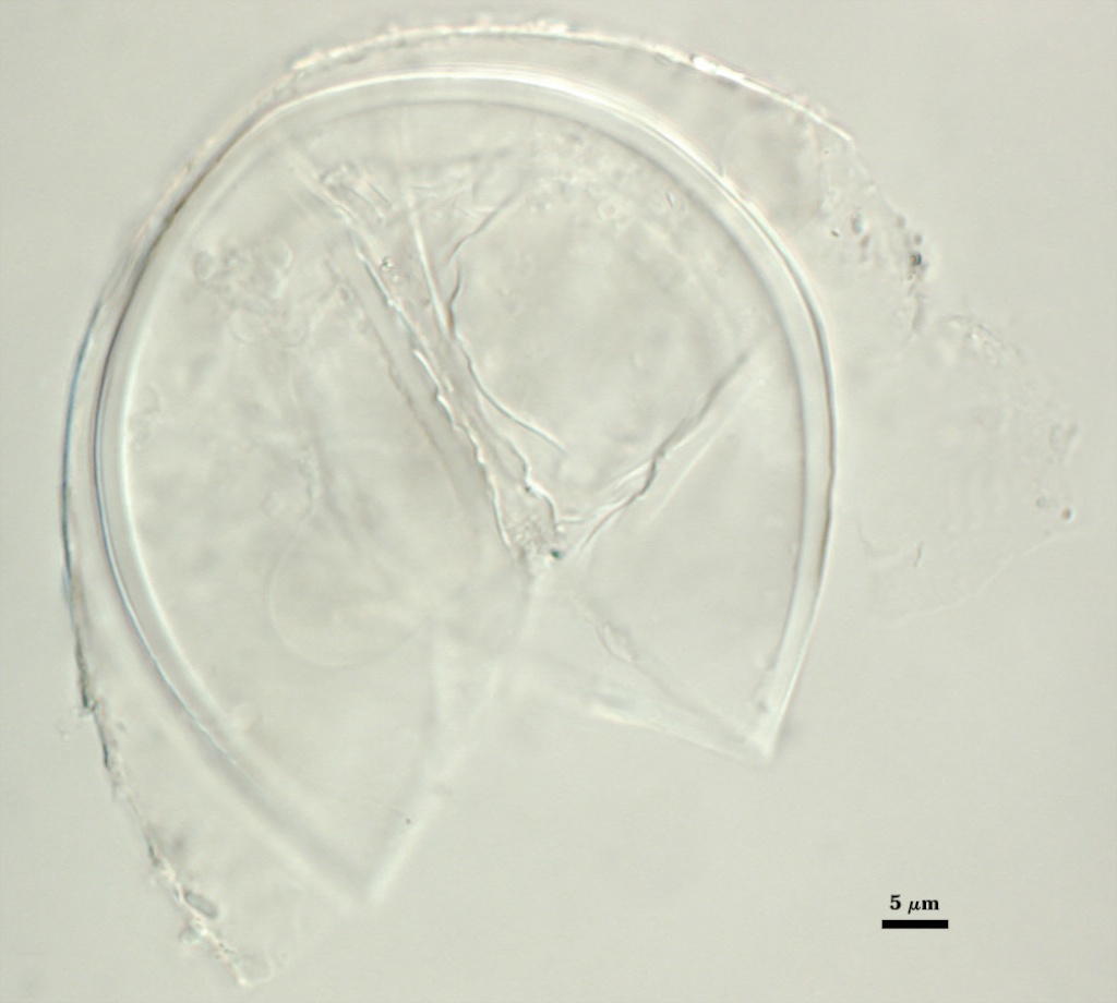
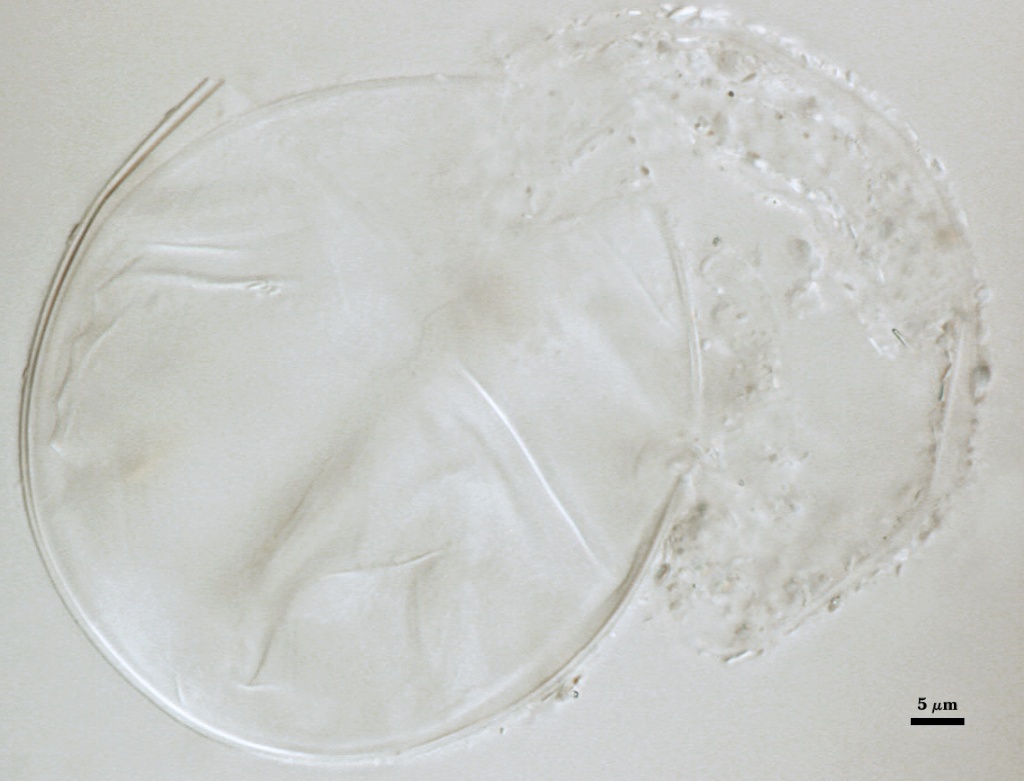
SPORE WALL: Consisting of three hyaline layers (L1, L2 and L3), all of which exhibit some flexibility when broken with applied pressure in a slide mountant.
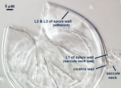
L1: A thin hyaline layer, < 1 µm thick, which is continuous with the wall of the neck of the sporiferous saccule. This layer often separates somewhat from the inner two layers.
L2: A thin hyaline layer, < 1 µm thick, which usually is adherent to L3. It is recognizable as a distinct layer by a distinct inner margin and also when it occasionally separates from L3.
L3: A thicker hyaline layer than either L1 or L2, ranging from 1.3-4 µm thick, which together with L2 form an endospore which separates the spore contents from that of the saccule neck.
| In PVLG | ||
|---|---|---|
 |  |  |
| In 1:1 v/v PVLG and Melzer’s reagent | ||
|---|---|---|
 |  |  |
Cicatrix
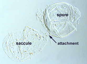
Scar indicating region of contact between spore and saccule neck during spore synthesis; diameter of 6-8 µm (mean = 7.2 µm ). The wall of the cicatrix is a slightly upraised region on the wall of the “endospore” formed by L2 and L3 of the spore wall.
Sporiferous Saccule
COLOR: Hyaline.
SHAPE: Oblong
SIZE DISTRIBUTION: 40-72 µm, mean = 53 µm.
SACCULE WALL: One layer with an outer coating of granular material, < 0.5-1.0 µm.
DISTANCE FROM SACCULE TO SPORE: 10-30 µm.
 |  |
Mycorrhizae
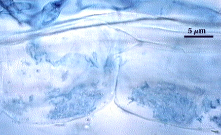
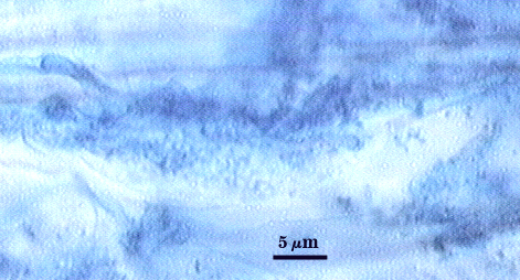
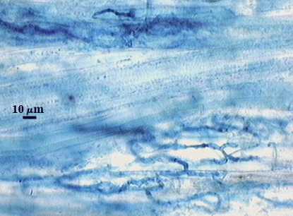
Arbuscules in corn mycorrhizae are very difficult to see because they stain so lightly in trypan blue. Coiled and straight hyphae in entry points widely scattered on roots stain more darkly and so are readily detectable. Coiling most most extensive near entry points, but the hyphae of an “infection unit” (all hyphae originating from an entry point) remain fairly localized with little overlapping between units. Intraradical hyphae are 2-3 µm wide. Contrast of photos below are enhanced digitally to see structures more clearly.
| Arbuscules in corn roots | |
|---|---|
 |  |
| Entry points & intraradical hyphae in corn | |
|---|---|
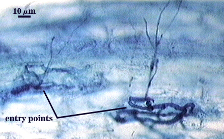 |  |
Notes
When spores float in water after extraction, the saccule often is oriented downward so that it is not always easy to detect. Saccules generally are 1.5 times as long as they are wide (e.g. 42×60 µm, 40×65 µm, 48×70 µm). When saccule remnants (resembling subtending hyphae) are present, spores are almost impossible to distinguish from Paraglomus occultum. However, spores tend to be more consistently globose in shape and many spores in any extractable spore population are likely to be either sessile or have saccules attached (the tipoff).
When this species was classified as a member of Acaulosporaceae, it was considered atypical because spores do not contain any germinal walls. The innermost layer of the spore wall is flexible, but shows no evidence of forming a “germination orb” nor is it bi-layered. Instead, spore wall structure is similar to that of other acaulosporoid species in Archaeospora.
High Resolution Images | |
|---|---|
 |  |
 |  |