Acaulospora denticulata
(reference accession CL139)
Whole Spores
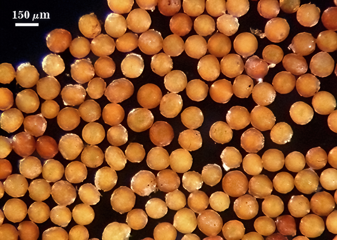
 COLOR: Pale orange-brown (0-20-80-0) to dark orange-brown (0-50-100-0), with most of intermediate color (0-40-100-0).
SHAPE: Globose, subglobose.
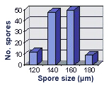
SIZE DISTRIBUTION: 120-180 µm, mean = 149.3 µm (n=120).
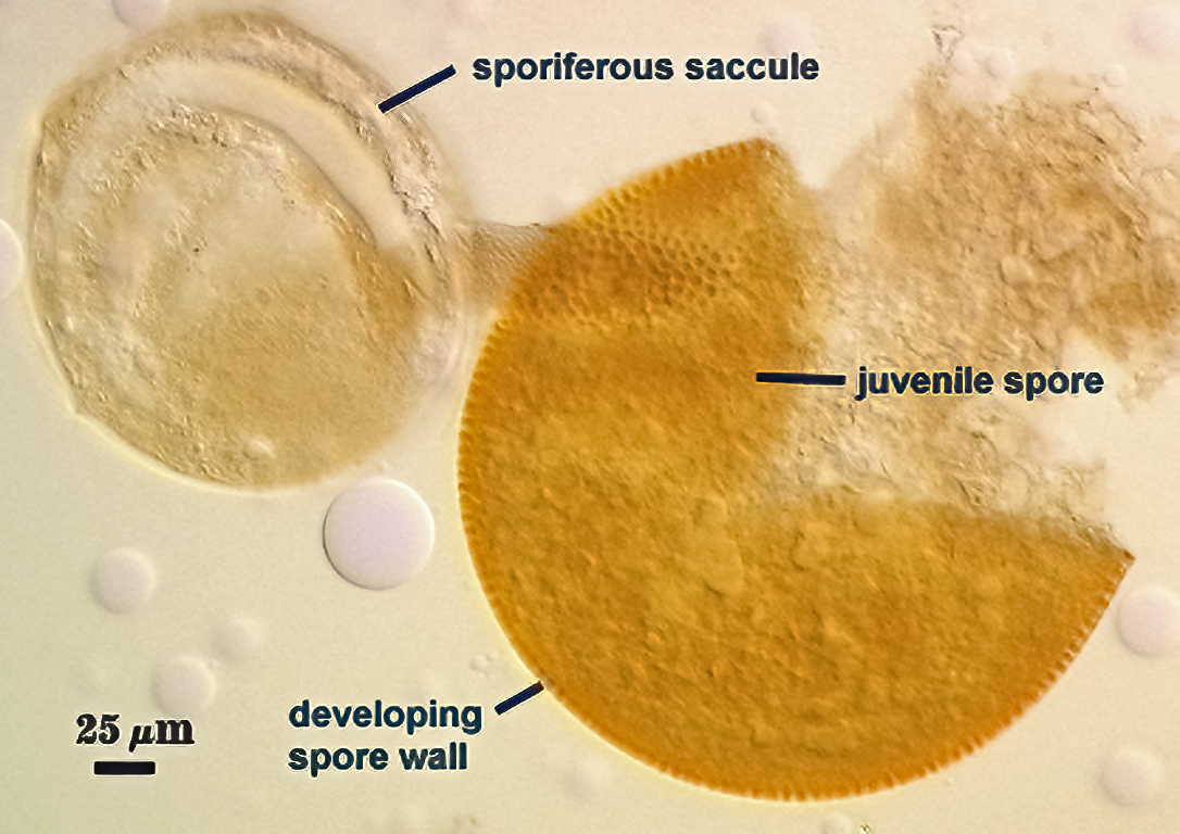
Subcellular Structure of Spores
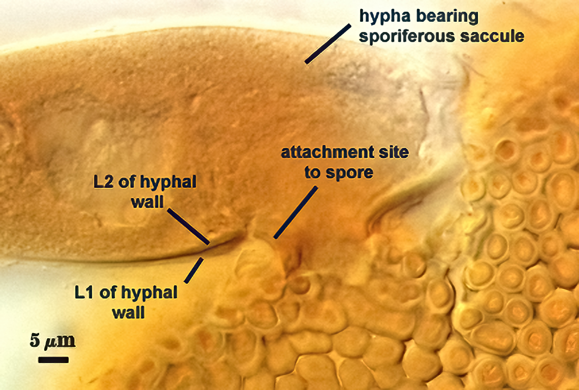
SPORE WALL: Three layers (L1, L2, and L3), the outer one continous with the wall of the neck of the parent sporiferous saccule (left photo) and the latter two being synthesized as soon as the spore wall begins to form (right photo).
| Differentiation of "denticulate" ornamentations as part of L2 spore wall | |||
|---|---|---|---|
|
|
|
|
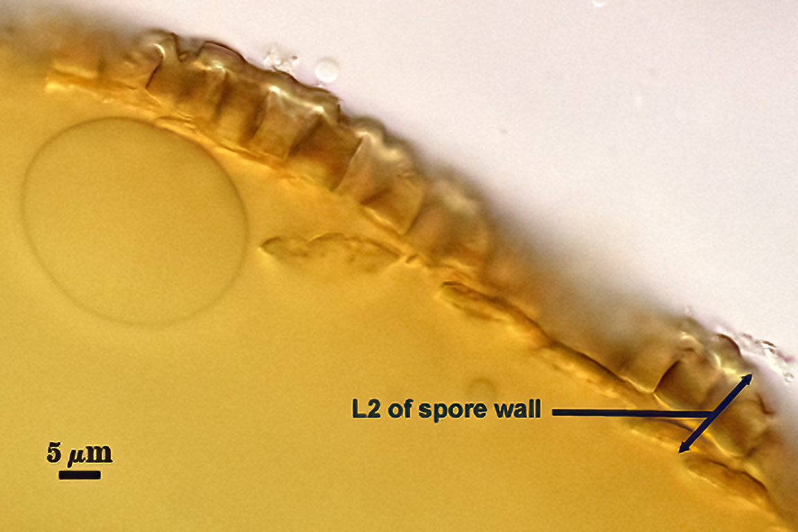
L1: Hyaline, 0.6-1.6 µm thick, degrading and sloughing early in spore wall differentiation and thus often are absent even before ornamentations on the laminate layer (L2 below) are fully formed. When present, usually adherent to developing ornamentations of L2.
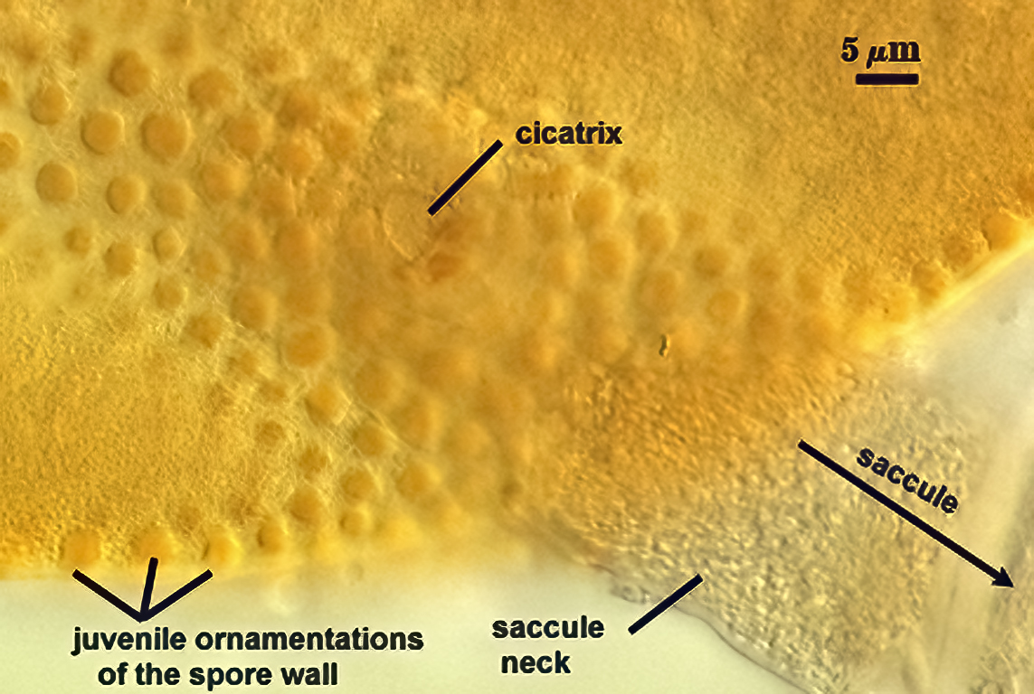
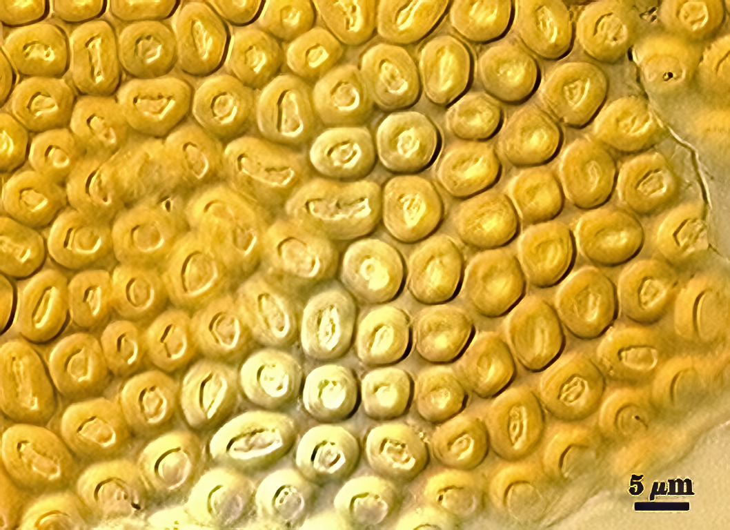
L2: Originates as a single layer 0.6-0.8 µm thick with stubby yellow-brown (0-20-100-0) “knobs” attached that are 2.2-2.6 µm wide and 3.0-3.2 µm long (the width of the spore wall at this juncture). Circular to oblong projections arise from the “knobs” and have a smooth outer edge or form a polygon of six sides. Circular projections are 4.2-6.8 µm wide (most 4-5 µm), whereas oblong projections can be as long as 9 µm. Each projection has a center cavity, 1.25-2.6 µm wide and 0.6-1.5 µm deep surrounded by a lip 1.25-1.5 µm wide. This layer is organized differently from a laminate layer in that it has a basal sublayer and the remaining structure results from organization of the ornamentations. Composite thickness ranges from 2.8-8.6 µm. At maturity, the pore between spore and saccule neck is closed by thickening of the sublayer (minus dentations) and becomes an “endospore” (completely enclosing the spore contents).
L3: A single hyaline layer, rarely thicher than 0.8 µm, which is detectable only when it separates from the spore wall. In vigorously crushed spores, this layer separates enough from the spore wall to appear as a separate flexible inner wall. It is not treated as one (despite similarity in appearance) because of close association with the spore wall analagous to a layer of similar position in many other Acaulospora species (e.g. A. scrobiculata, A. mellea, A. tuberculata).
| Spores mounted in PVLG | |||
|---|---|---|---|
|
|
|
|
| Spores mounted in PVLG and Melzer’s reagent (1:1 v/v) | |||
|---|---|---|---|
|
|
|
|
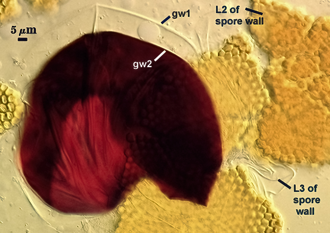
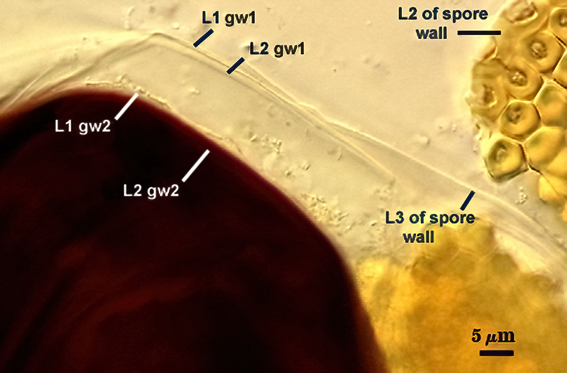
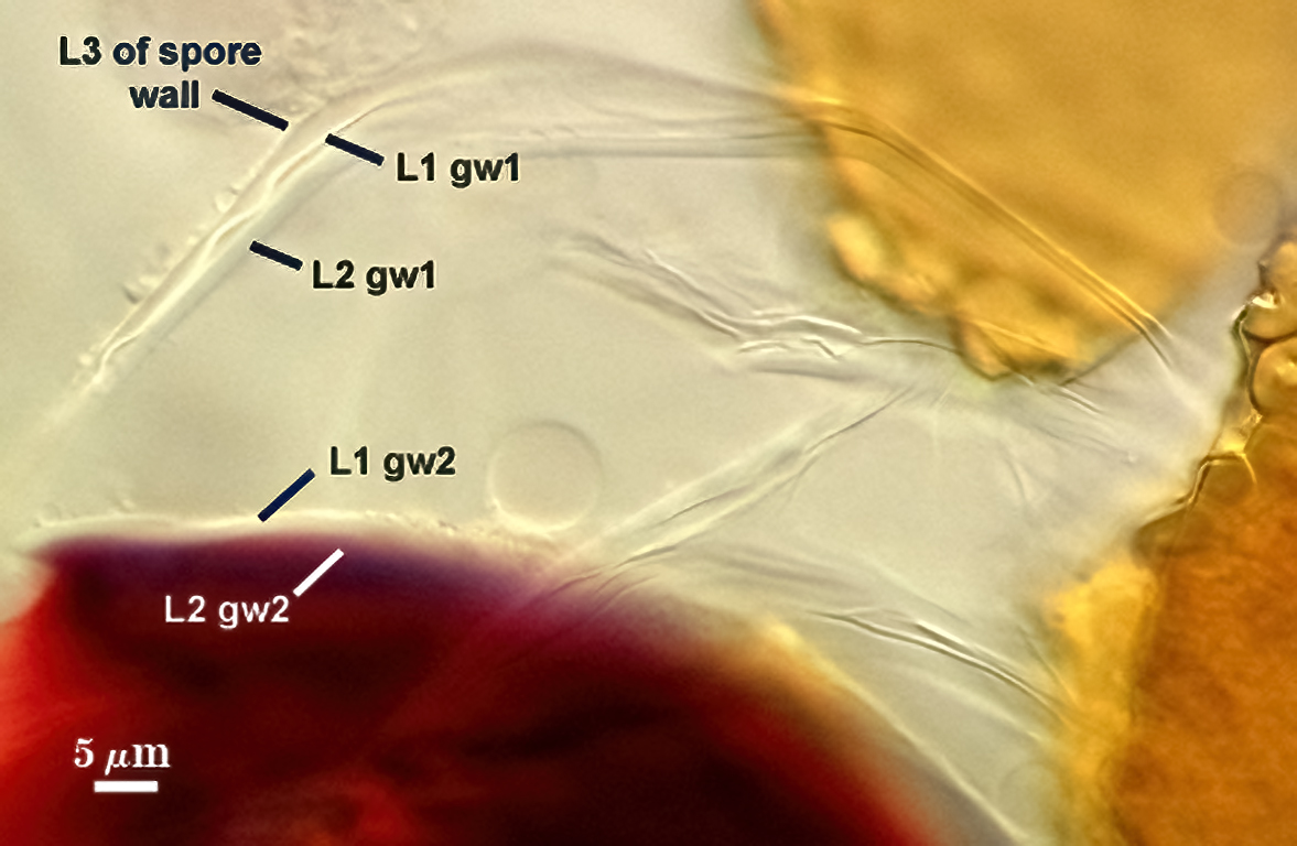
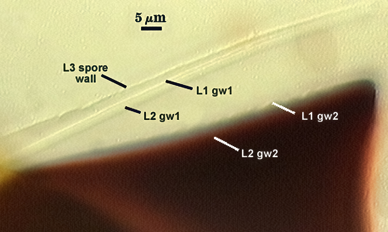
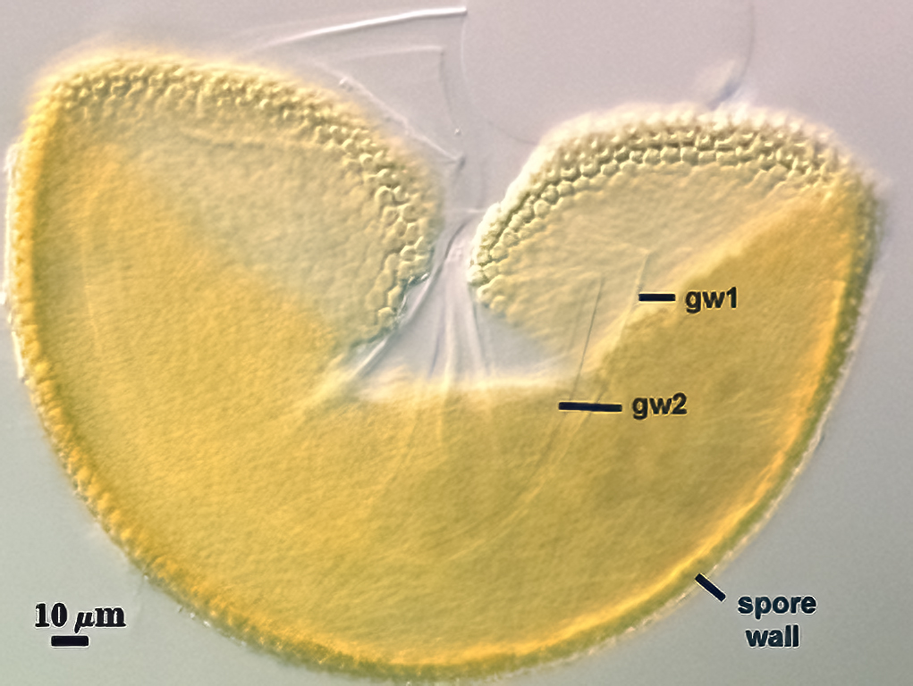
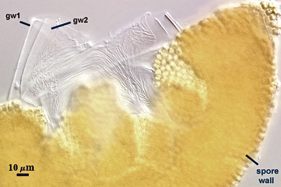
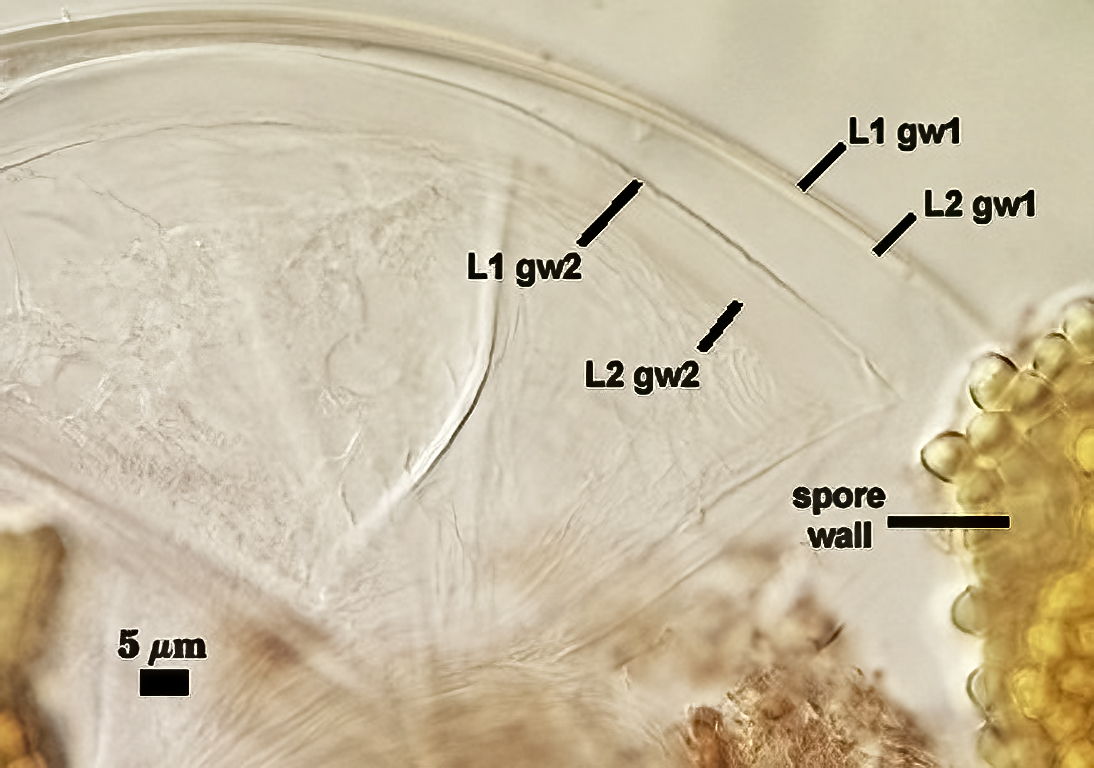
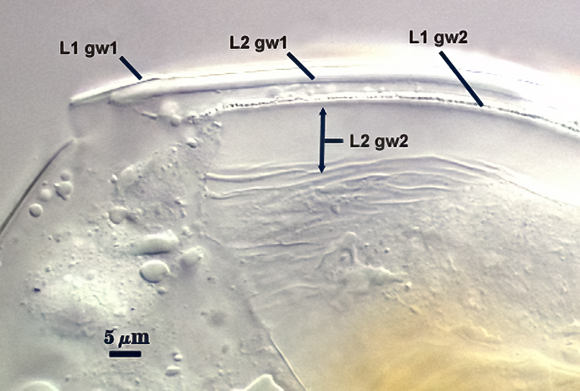
GERMINAL WALLS: Two hyaline flexible inner walls (gw1 and gw2) are synthesized sequentially after the spore wall has completed differentiation.
GW1: Two layers are formed (L1 and L2). L1 is 0.4-0.6 µm thick and in close proximity usually to L2, which is 1.2-1.4 µm thick. This wall usually is easy to see because it separates readily from the spore wall and iw2.
GW2: Two adherent layers are formed (L1 and L2). L1 is 0.6-1.0 µm thick, with granular excresences (or “beads”) that tend to become dislodged and float away with applied pressure. These “beads” are stabilized after preservation in formalin, but otherwise may be absent on mounted spores within a few months of storage. L2 is plastic enough that it has been termed “amorphous”. It is 3-10 µm thick in PVLG-based mountants, depending on amount of pressure applied to it while breaking the spore; staining red-purple (20-80-20-0) to dark red-purple (40-80-60-0) in Melzer’s reagent.
Cicatrix
A circular to ovoid scar indicating region of contact between spore and saccule neck during spore synthesis; it consists of denticulate ornamentations packed around a relatively smooth depression, 6.0-11.3 µm in diameter (mean = 9.4 µm ) at its widest. The connection between saccule neck and spore consists of only the L1 layer of the spore (continuous with the wall of the saccule neck) initially, followed by synthesis of sublayers of the L2 (laminate) layer which form a lip 2.4-3.5 µm high between saccule neck and spore.
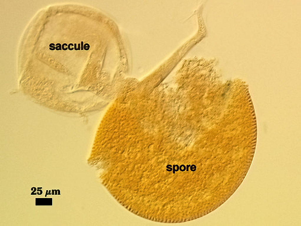
Sporiferous Saccule
COLOR: Hyaline.
SHAPE: Mostly globose (see photos above).
SIZE DISTRIBUTION: 130-160 µm, mean = 151 µm.
COMPOSITE WALL THICKNESS: 1.4-4.2 µm.
SACCULE WALL: One layer, smooth surface.
DISTANCE FROM SACCULE TO SPORE: 60-80 µm, rarely 100 µm.
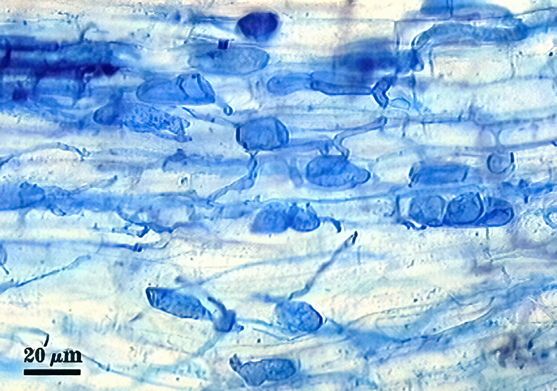
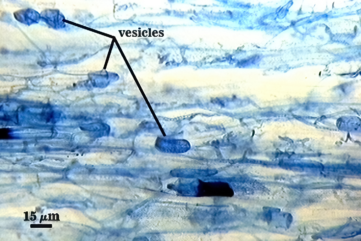
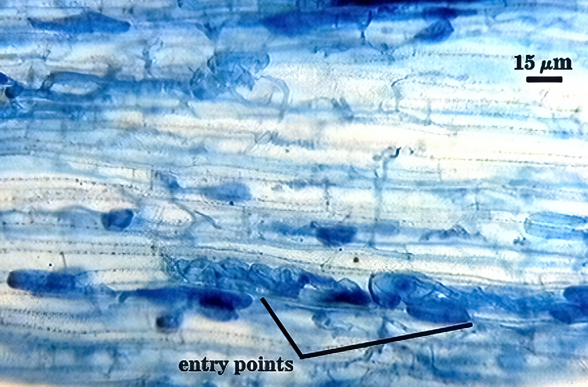
Mycorrhizae
Intraradical arbuscules tend to stain lightly in trypan blue, whereas intraradical hyphae and vesicles are more variable, from light to darkly stained. Hyphae are coiled mostly near entry points, 3-6 µm in diameter; otherwise thinner (2-4 µm) and usually growing parallel to each other and interconnecting via branches that form “H” and “h” connections. Vesicles often are localized in clusters that are patchily distributed along a mycorrhiza. Many appear to form near entry points (primary or secondary ingress) and many are lobed or irregularly-shaped.
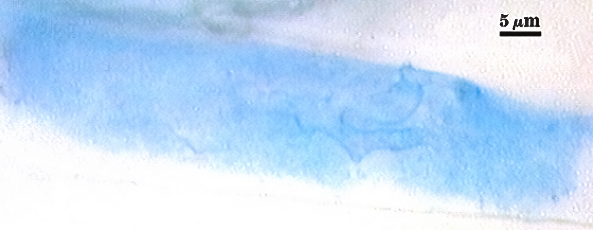
| Lightly staining arbuscules in corn cortical cells | |
|---|---|
|
|
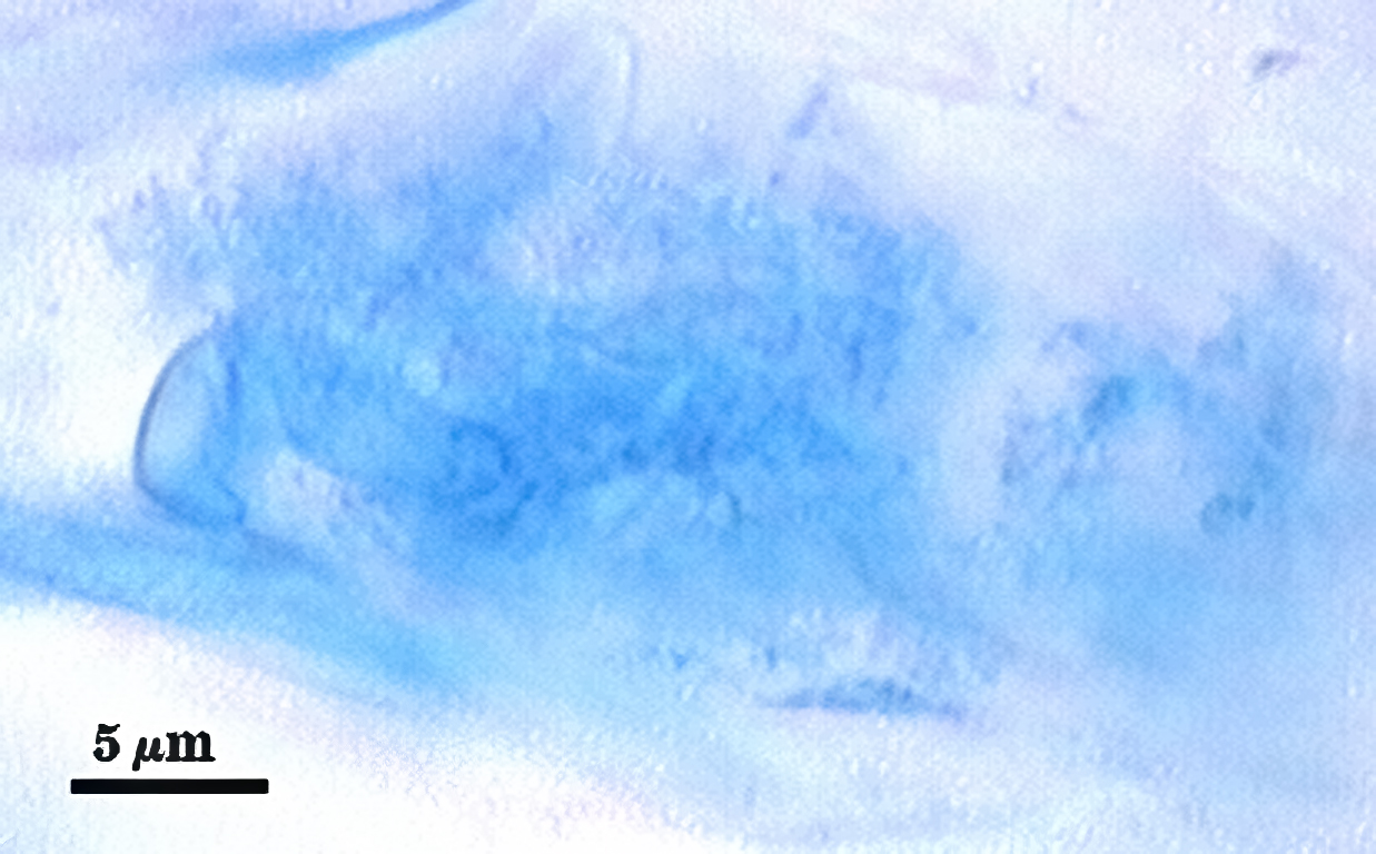
| Dark-staining mycorrhiza in corn, showing all structures | ||
|---|---|---|
|
|
|
Germination
An ovoid “germination orb” is presumed to form on gw2 like that found in other Acaulospora species, but it has not yet been observed in specimens examined.
Notes
Immature spores and saccules are the same salmon color (0-20-60-0). Differentiation of dentations is almost completed before contents of the saccule are emptied and always before the saccule has detached.