Acaulospora foveata
(reference accession BR861)
Whole Spores
| Spore Growth | |
|---|---|
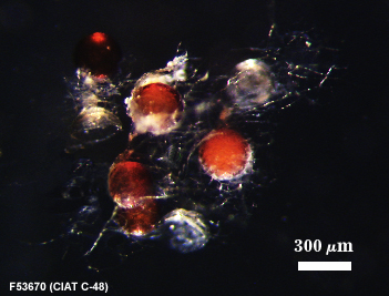 | 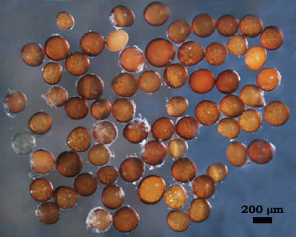 |
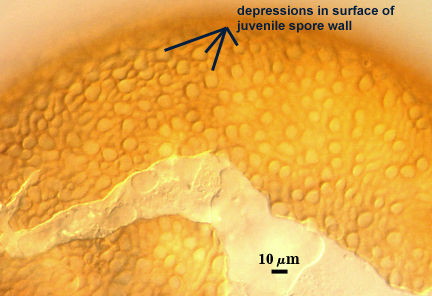
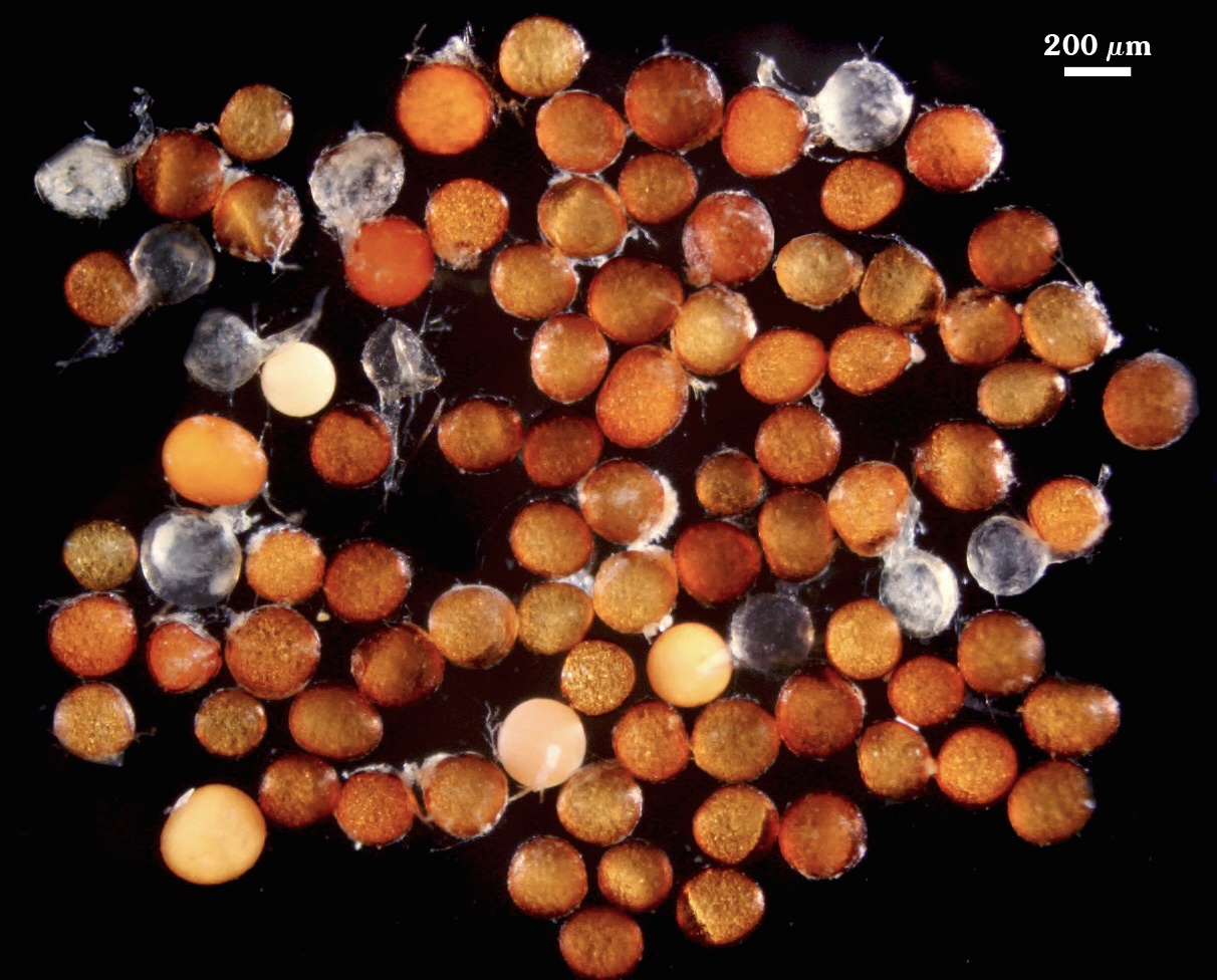
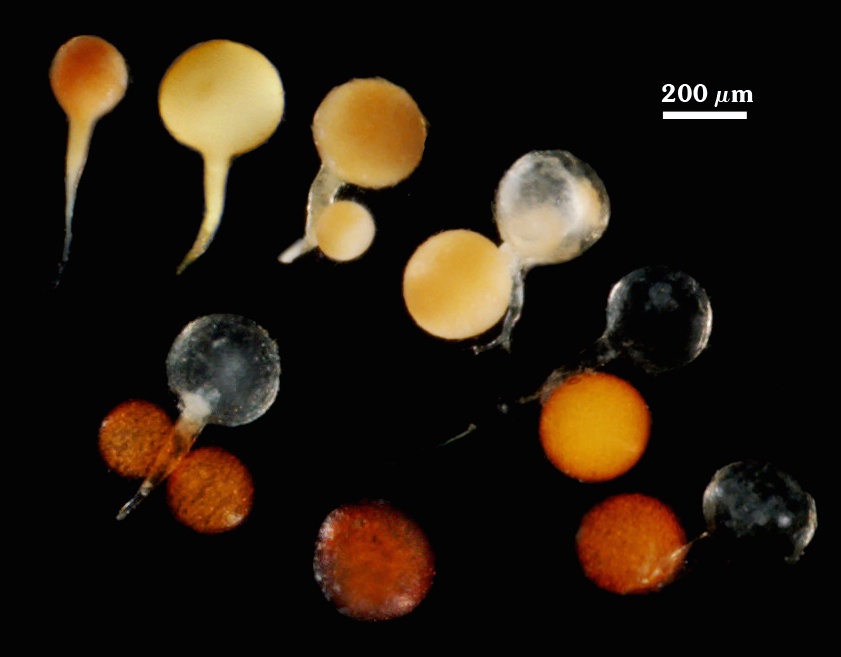
 COLOR: Red-orange (0-50-60-5) to dark red- brown (40-50-70-10). Immature spores initially are cream-colored and gradually acquire an orange tint as the spore wall begins to differentiate (see photo).
SHAPE: Globose to subglobose, sometimes irregular.
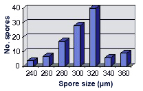
SIZE DISTRIBUTION: 240-360 µm (mean = 289 µm, n = 58).
Subcellular Structure of Spores
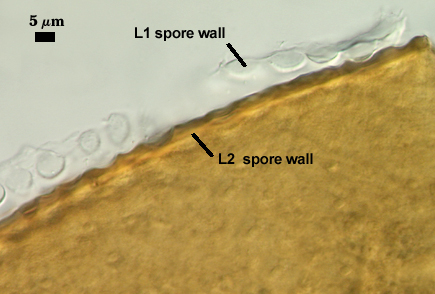
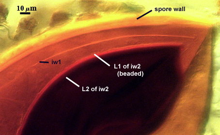
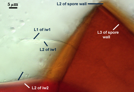
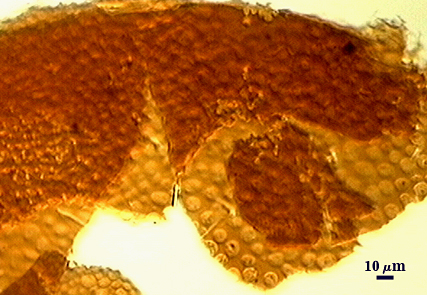
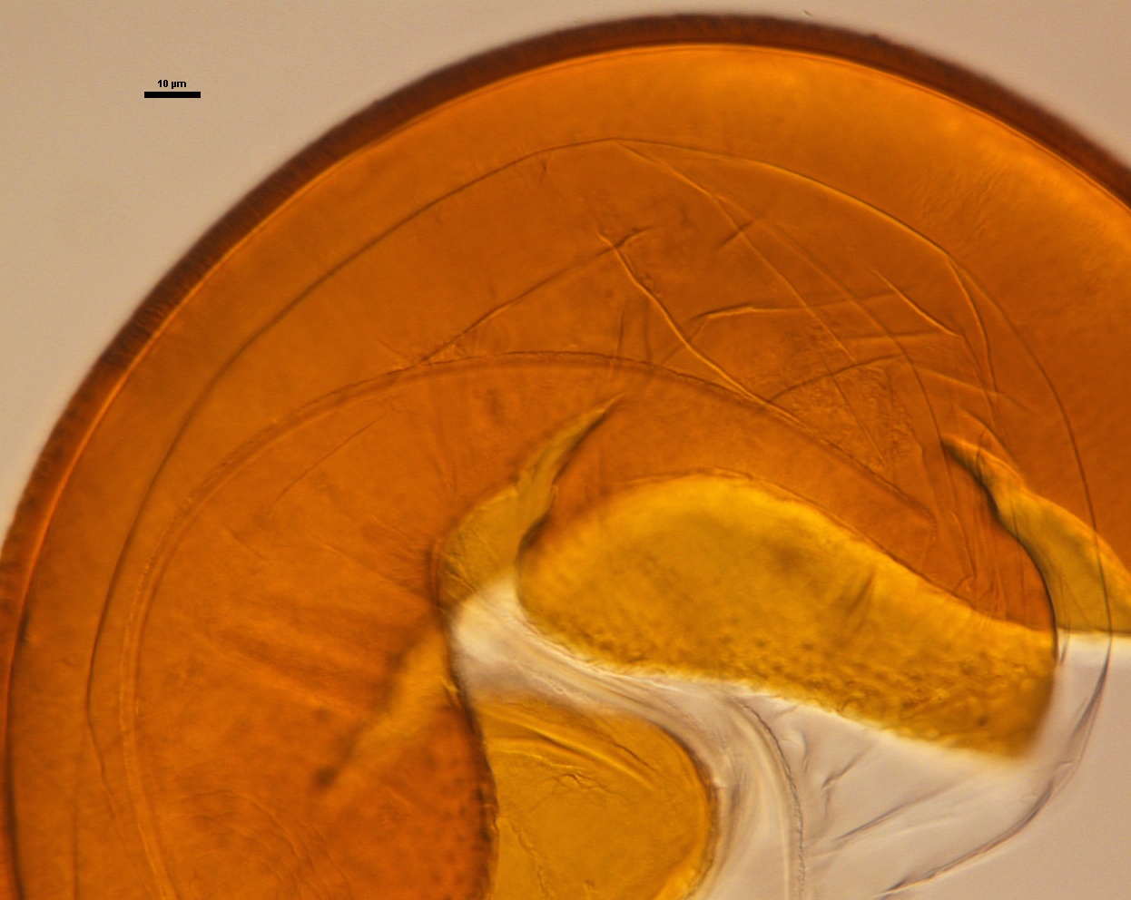
SPORE WALL: Three layers (L1, L2 and L3) are usually evident, with the outer layer continous with the wall of the neck of the parent sporiferous saccule. The inner two layers are synthesized sequentially as the spore forms on the neck of the saccule. A third layer homologous with that in spores of other large ornamented species (e.g., A. tuberculata).
| Foveata Spore Structure | ||||
|---|---|---|---|---|
|
|
|
|
|
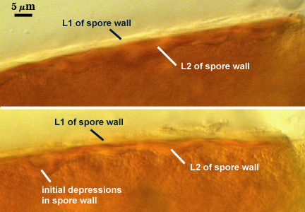
L1: Hyaline, 2-2.4 µm thick; degrading and sloughing with collapse and dehiscence of the sporiferous saccule (see first photo above).
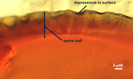
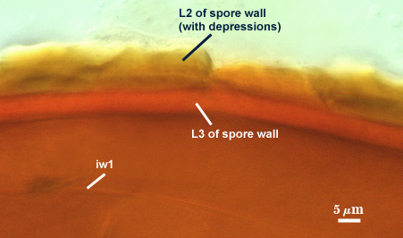
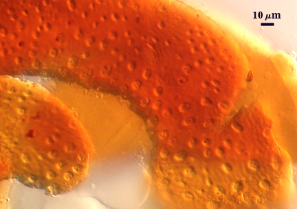
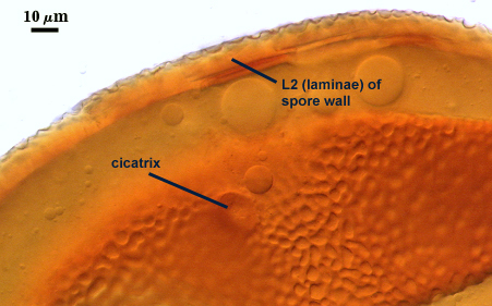
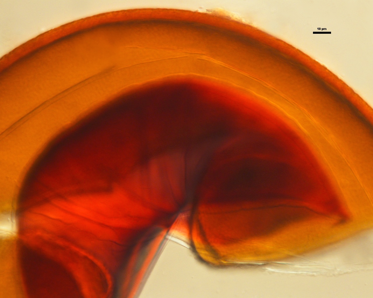
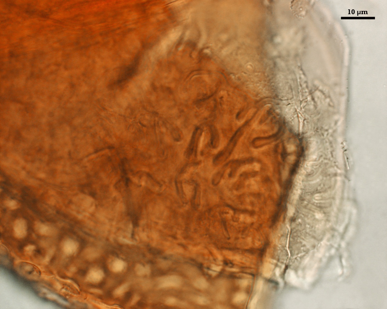
L2: A layer that thickens with synthesis of orange-red sublayers (or laminae); 9-18 µm (mean = 14.6 µm ) thick at maturity. Initially a single layer, 2-4 µm thick, forms in undulations that establish the shape and depth of surface concave depressions (or pits), with additional sublayers forming horizontally beneath to establish a an underlying base (see photos above). Depressions are circular to ovoid, 8-12 µm across and 0.5-3 µm deep in the mature layer. This layer does not produce any reaction in Melzer’s reagent.
L3: A layer that forms with synthesis of additional concolorous sublayers that are not readily distinguishable in PVLG, but more evident in Melzer’s reagent because it produces a dark red-brown reaction. This layer often is fused with L2, but sometimes separates (see photo above), 3-5 µm thick when measurable. This layer may be homologous with an inner separate laminate layer (L3) in spores of A. koskei and A. laevis.
| Spores mounted in PVLG | |
|---|---|
|
|
| Spores in PVLG & Melzer’s reagent | |||
|---|---|---|---|
|
|
|
|
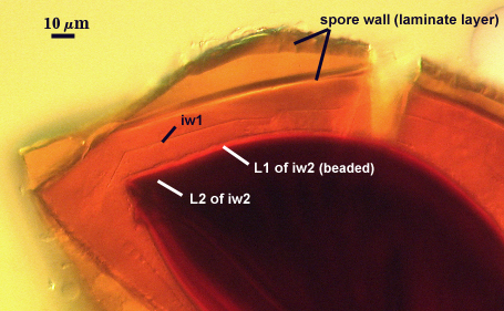
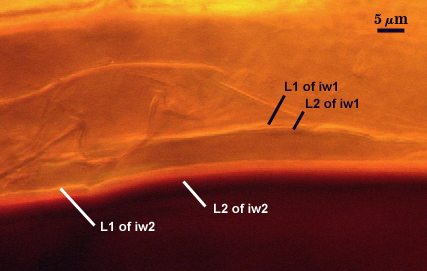
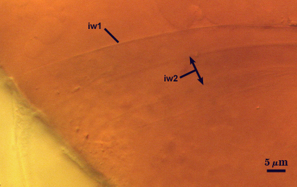
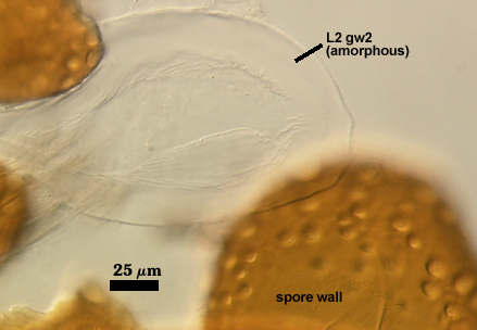
GERMINAL WALLS: Two hyaline flexible inner walls (gw1 and gw2) are present. They can be seen only after spores have completely cleared when mounted in PVLG or Melzer’s reagent, which can take as long as 30 days.
GW1: Consisting of two adherent layers (L1 and L2) are formed, both are of near-equal thickness, ranging from 0.6-1.0 µm thick. This wall forms completely independent of the spore wall and thus has no direct developmental connection with it.
GW2: Consisting of two adherent layers (L1 and L2) are formed that together are 5-12 µm when measured in PVLG. L1 is hyaline, 1.5-2.2 µm thick with granular excresences (or “beads”) that tend to become dislodged and float away when pressure is applied to it. These “beads” are stabilized after preservation in formalin, but otherwise may be absent on mounted spores within a few months of storage. L2 is hyaline and somewhat plastic (probably equivalent to a “coriaceous” wall using traditional terminology). It is 2-10 µm thick in PVLG-based mountants, staining pinkish red (20-80-60-0) to a reddish-purple (40-80-60-0) in Melzer’s reagent.
Cicatrix
A circular to ovoid scar indicating region of contact between spore and saccule neck; it consists of closely packed tubercles surrounding an unornamented depression, 12-16 µm in diameter (mean = 13.8 µm) at its widest.
Sporiferous Saccule
| Isotype | INVAM |
|---|---|
|
|
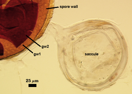
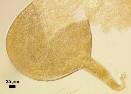
COLOR: White to cream when immature, hyaline when emptied of contents.
SHAPE: Mostly globose to subglobose.
SIZE: 260-320 µm, mean = 282 µm
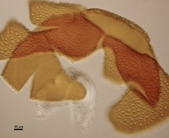
SACCULE WALL: A single hyaline layer with fine sublayers; the outermost sublayers appearing somewhat flakey; 3.5-4 µm thick. In the isotype (see left photo above), flakiness of the saccule surface was more pronounced and the wall was thicker (4-10 µm).
SACCULE NECK: Tapering from the saccule to the region of spore attachment; saccule neck wall hyaline and 1.8-2.2 µm thick
DISTANCE FROM SACCULE TO SPORE: Not measured in spores examined thus far.
Notes
The description of this species (Janos and Trappe, 1982) was based on field-collected spores, many of which appeared to be parasitized by other fungi (see left photo). None of the type specimens contained inner (germinal) walls (see right photo). In fact, the authors only description of such structures is the statement ” .....an adherent but separable hyaline inner layer 3 µm thick”. It is likely, from this measurement, that both gw1 and gw2 were fused together.
The accession from which live spores were obtained is a trap culture and therefore contains more than one species. For that reason, we cannot report mycorrhizal morphology at this time. Attempts to obtain a monospecific culture from extracted spores so far have failed. We continuously maintain the trap culture by reseeding with the expectation of increasing numbers of healthy spores.
Janos and Trappe
| Spore Growth | |
|---|---|
|
|
High Resolution Images | |||
|---|---|---|---|
 |  | ||
 |  | ||
 |  | ||
Reference
- Janos, D. P. and J. M. Trappe. 1982. Two new Acaulospora species from tropical America. Mycotaxon 15: 515-522.