Glomeraceae - Piroz. & Dalpé emend C. Walker & Schüßler
Spores Glomoid Definition
Spores glomoid, produced at or near the soil surface, in sporocarps with or without a peridium or as spores singly or in clusters. A diagnostic (but fairly degenerate) sequence from the SSUgene is: TGTYADGGCAYYRCACYGG.
All species formerly classified as members of Glomus are included except for those now grouped in the families Claroideoglomeraceae, Diversisporaceae or Paraglomeraceae.
Vegetative (Mycorrhizal) Features
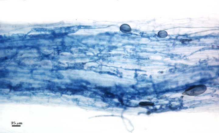
Intracellular Arbuscules generally stain darkly (using trypan blue on specimens of this website). This property can be somewhat variable within the same root or between roots, depending on homogeneity of stain diffusion and reactivity, but structures all exhibit relatively high contrast.
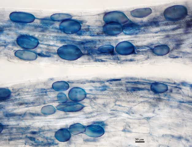
Intraradical Vesicles may or may not be formed, depending on fungus and environment during mycorrhizal development in a host plant. When abundant, vesicles may be localized near entry points but more often are widely dispersed throughout a mycorrhiza. They generally are oblong to elliptical in shape and usually stain darkly in trypan blue and other stains. They usually form late in mycorrhizal development (in pot cultures) or more likely within field roots as they lose nutrient absorption capability and undergo senescence.
Intraradical hyphae may branch at acute or oblique angles or be coiled in entry point regions. Colonization hyphae usually grow parallel to each other and to the root axis, generally vary from 1.5 to 4 µm wide, and interconnect via right-angle or acute-angle branches (often referred to as “H connections”). They usually stain darkly in trypan blue and other stains.
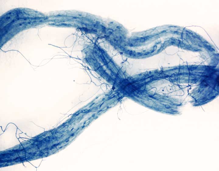
Extraradical hyphae varying considerably in abundance, distribution, and morphology among species. Some are very fine (thin) whereas others are very robust (wide) (e.g., Rhizophagus clarus).
| Arbuscules, Vesicles and Hyphae | |||
|---|---|---|---|
 |  |  |  |
Asexual Spores
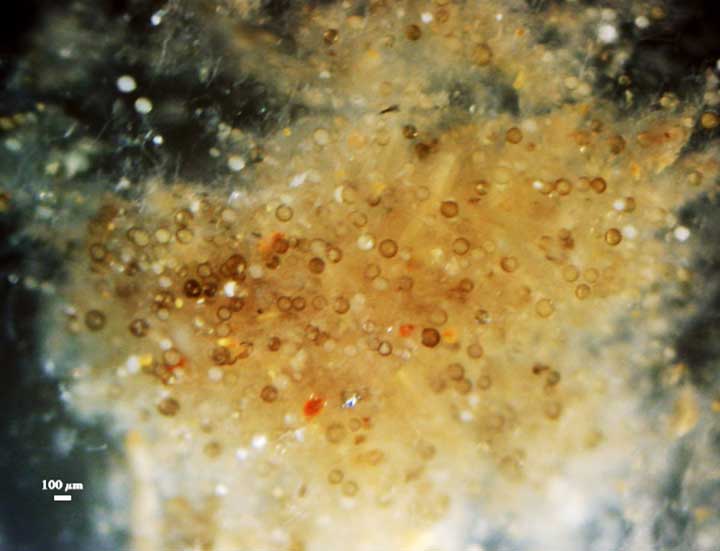
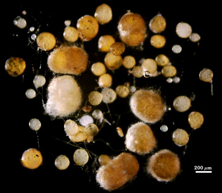
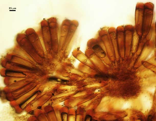
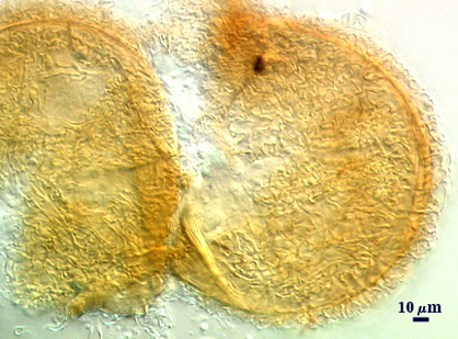
Spores are produced singly, in aggregates, in an unorganized hyphal matrix, or in a highly ordered hyphal matrix (see photos below), with layers of the spore wall usually continuous with a wall of the subtending hypha. They also may form within roots, possibly as a substitute or as a replacement for vesicle development in some species. In the table below (left) is a photo of R. intraradices spores amassed near a root. Below the second image is of F. mosseae that forms spores both singly without a perineum and final image (right) is in small clusters where spores are bound together by peridial hyphae and an organized sporocarp of G. clavisporum with spores formed around a central hyphal plexus.
| F. mosseae | ||
|---|---|---|
|
|
|
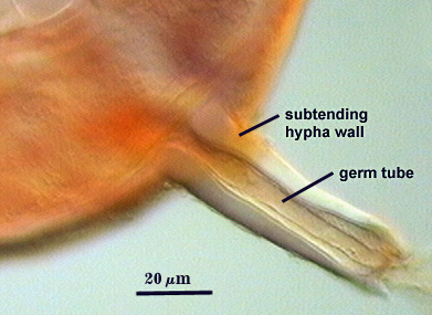
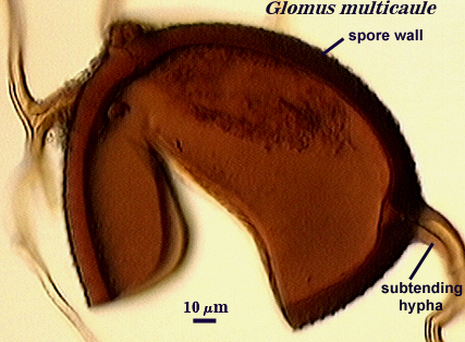
Develop on a cylindrical, flared, or constricted unspecialized fertile hypha. In most cases, spores are borne terminally, but in rare cases may form appear to form intercalarily because of hyphae at opposing poles (see the first image below for a voucher specimen of G. multicaule).
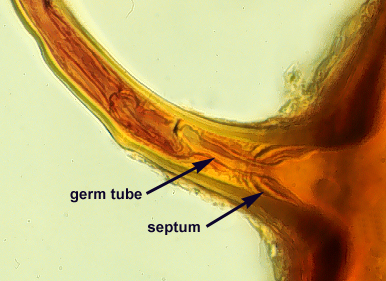
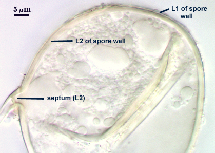
Partition contents from that of the subtending hypha by different structural mechanisms such as an amorphous plug, a septum, an inner sublayer of the laminate layer of the spore wall, or thickening of all sublayers of the laminate layer of the spore wall.
| Subtending hypha | |||
|---|---|---|---|
|
|
|
|
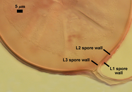
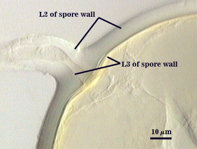
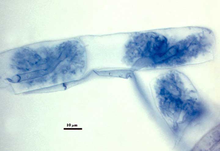
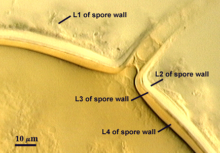
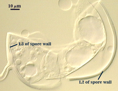
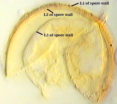
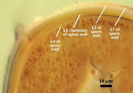
Form the greatest number of layers with the widest variety of phenotypes amongst the three families of Glomales. Only a few species do not have an evanescent mucilagenous outer layer (stains pink to red in Melzer’s reagent) that is a major component of the juvenile spore and hyphal walls. All have a rigid layer with sublayers (or laminae) that is continuous with the subtending hyphal wall.
| Phenotypes | ||
|---|---|---|
|
|
|
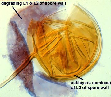
May synthesize a separable thin (and hence flexible) sublayer of the laminate layer of the spore wall which previously has been defined as a “membranous wall”. However, it originates as part of the subtending hypha and remains attached in some broken spores, so it is not a separate wall.
| Membranous wall | ||
|---|---|---|
|
|
|
Germinate usually by emergence of the germ tube through the lumen of the subtending hypha.
| Germinate | ||
|---|---|---|
|
|
|