Septoglomus viscosum
(reference accession MD215)
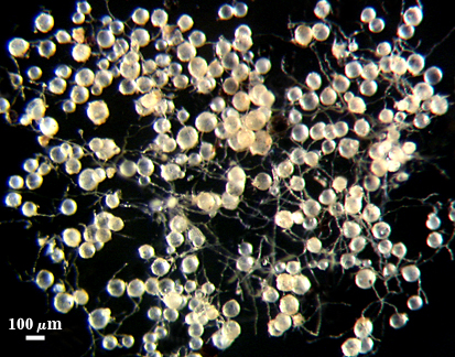
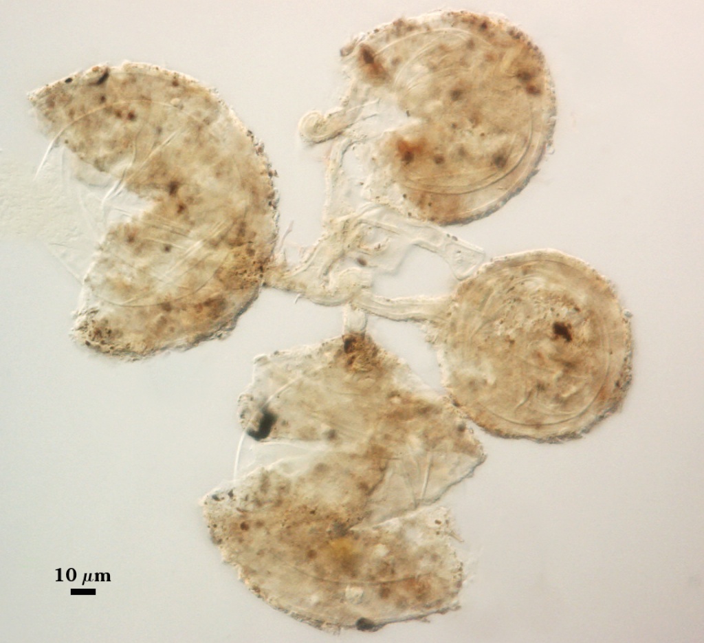
Whole Spores
 COLOR: Subhyaline (0-0-5-0) to pale straw (0-5-20-0) at darkest; surface of spores usually appears dull because of surface organic matter or debris.
SHAPE: Globose to subglobose, rarely irregular. Many spores are formed in loose aggregates branching from a common hypha.
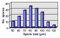
SIZE DISTRIBUTION: xx µm (mean = ). 50-120 µm (mean = 82 µm, n =160)
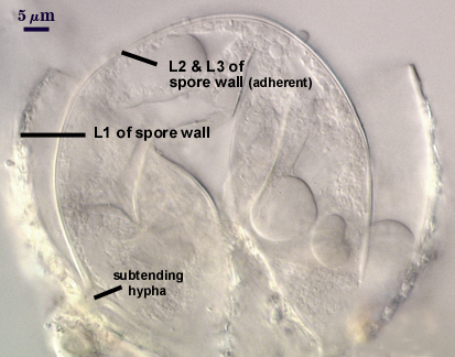
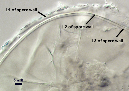
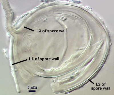
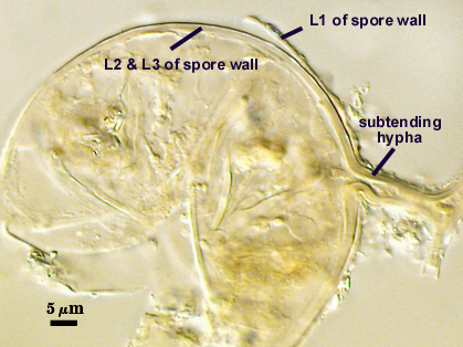
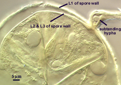
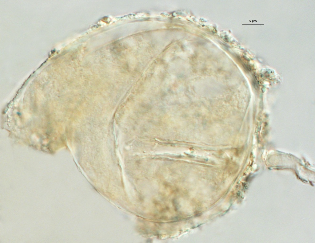
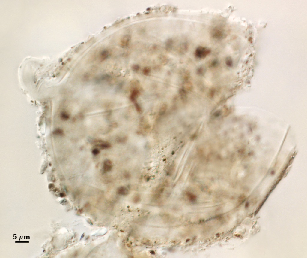
Subcellular Structure of Spores
SPORE WALL: Three layers (L1, L2, and L3), with the outer layer usually separating somewhat from the inner two layers (which usually are adherent).
L1: A semi-flexible hyaline layer; 1-2 µm thick when surface excretions or debris are absent, up to 5 µm thick with surface matter. This layer does not react in Melzer’s reagent, so the surface matter attached to L1 does not contain the carbohydrates of an outer mucilagenous layer in many other Glomus species (e.g., C. etunicatum, C. claroideum, or R. clarus). Energy dispersive spectrometric results and a Schiff periodic acid reducing solution test indicate this material is of soil rather than fungal origin (Walker et al., 1995). In aged spores, this layer tends to remain on the spore but tends to separate to varying degrees from both the subtending hyphal and spore wall.
L2: A thin semi-flexible hyaline layer (0.5 µm or less) with discrete edges so that it appears as a raised outer edge on L3 when tightly adherent. This layer was not recognized in the protologue of this species (Walker et al., 1995). It tends to separate from L3 in less than 10% of crushed spores, but is observed consistently (see photos above).
L3: A hyaline layer which thickens via sublayers (or laminae) during spore ontogenesis. This layer is more rigid than L1 or L2, but it has more flexibility than the brittle structural layers of many other glommed species. It also is not as flexible as the structural wall of some species (e.g., D. spurca) so that it produces folds on the inner surface after being crushed.
| In PVLG | ||
|---|---|---|
|
|
|
| In PVLG and Melzer's reagent | ||
|---|---|---|
|
|
|
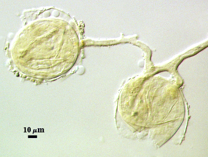
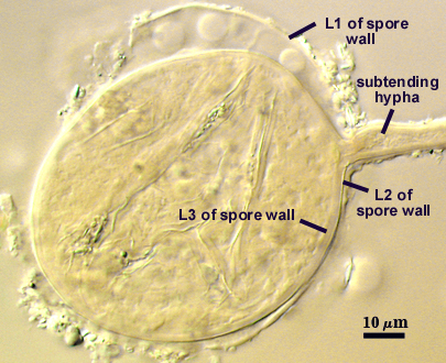
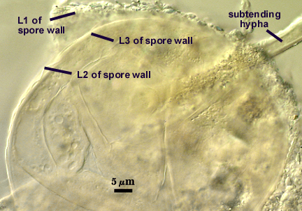
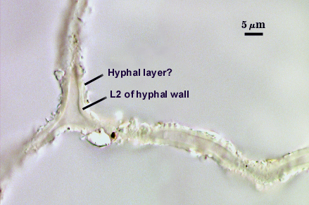
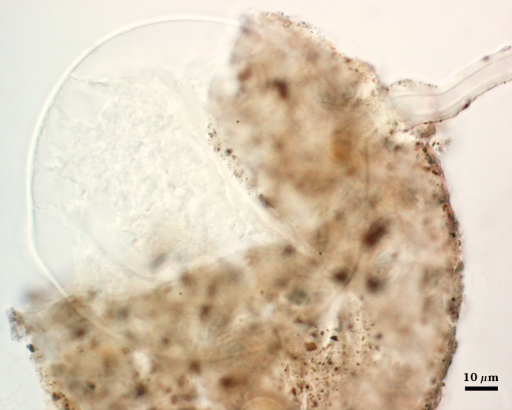
Subtending Hypha
SHAPE: Cylindrical to slightly flared, occasionally slightly constricted.
WIDTH: 8-11 µm at spore.
WALL STRUCTURE: Three hyaline layers (L1, L2, L3) in mature spores that are continuous with the three layers of the spore wall, although L1 and L2 become difficult to see (if they are present) in hyphae more than 100 µm from the spore. Hyphae in soil have a layer obviously continuous with L3 of the spore wall, but the outer matter is more granular and more likely to be of soil than fungal origin (Walker et al., 1995).
OCCLUSION: Appears to be absent in many spores. Sometimes, the spore wall thickens separate spore from hyphal contents (see photo below).
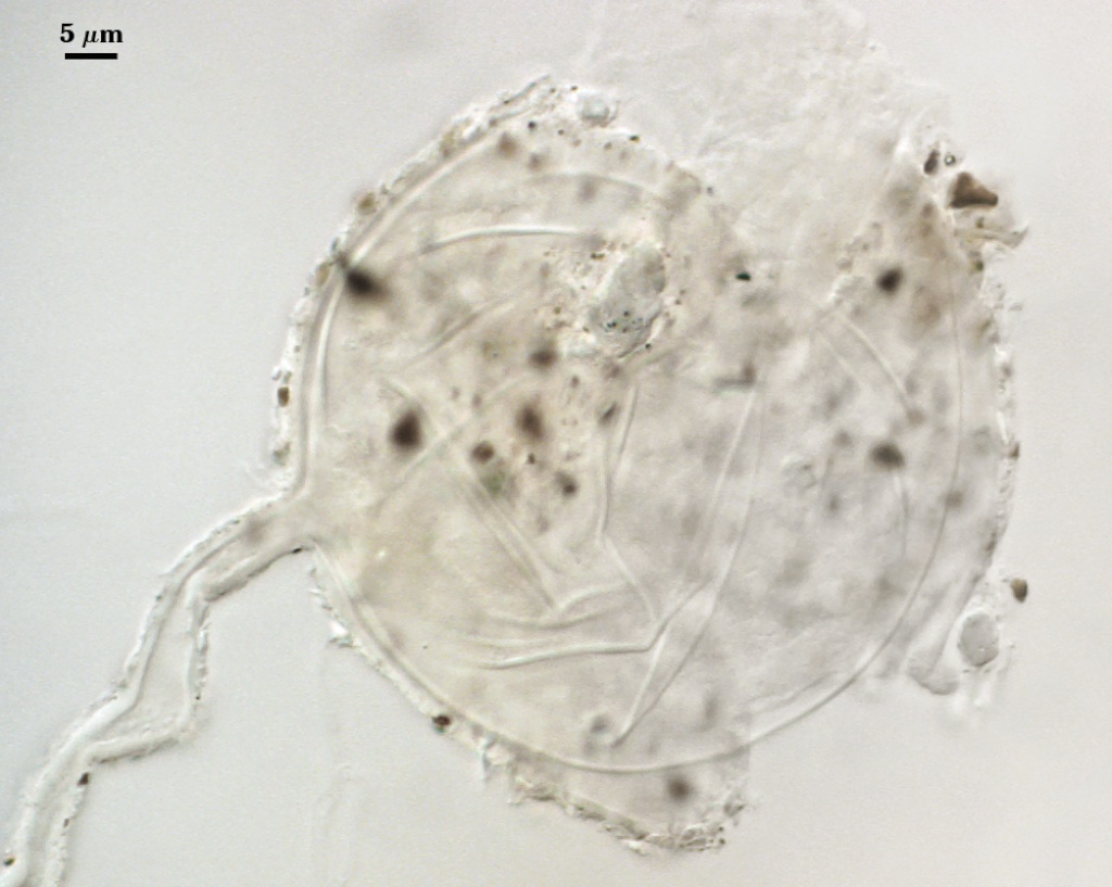
| Viscosum spore | ||
|---|---|---|
|
|
|
Notes
This isolate was obtained from Pat Millner (USDA-ARS Beltsville, MD, USA). It was easy to identify as S. viscosum because of the thorough and detailed description by Walker et al. (1995). The fungus is a prolific sporulator, with almost 50% of spores in culture organized in loose aggregates. It also produces abundant glomalin (Wright, pers. commun.). LSU sequences generated in our lab clearly place this species in the Rhizophagus clade. Similar evidence has been obtained from the SSU gene (Redecker et al., 2013)
Spores were somewhat larger than those of the type isolate of the species (44-97×46-94 µm, mean of 60 µm), but such variation in different spore populations is similar to that of other hyaline small-spored glomoid species such as Paraglomus occultum.
The images below can be uploaded into your browser by clicking on the thumbnail or can be downloaded to your computer by clicking on the link below each image. Please do not use these images for other than personal use without expressed permission from INVAM.
High Resolution Images | |
|---|---|
|  |
 |  |
 |  |
Links to Gene Sequences in Genbank
References
- Walker, C., M. Giovannetti, L. Avio, A. S. Citernesi, and T. H. Nicolson. 1995. A new fungal species forming arbuscular mycorrhizas: Glomus viscosum. Mycological Research 99:1500-1506.
- Redecker, D., A. Schüßler, H. Stockinger, S. Stürmer, J. Morton, and C. Walker. 2013. An evidence-based consensus for the classification of arbuscular mycorrhizal fungi (Glomeromycota). Mycorrhiza doi:10.1007/s00572-013-0486-y.